Engineering enzyme subsite specificity: preparation, kinetic characterization, and X-ray analysis at 2.0-A resolution of Val111Phe site-mutated calf chymosin.
Strop, P., Sedlacek, J., Stys, J., Kaderabkova, Z., Blaha, I., Pavlickova, L., Pohl, J., Fabry, M., Kostka, V., Newman, M.(1990) Biochemistry 29: 9863-9871
- PubMed: 2271625
- DOI: https://doi.org/10.1021/bi00494a016
- Primary Citation of Related Structures:
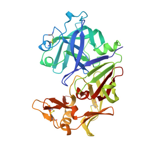
3CMS - PubMed Abstract:
Comparison of the three-dimensional structure of bovine chymosin with the structures of homologous aspartic proteinases complexed with peptide inhibitors shows that Val111 in chymosin occupies a position between the specificity subsites S1 and S3. A mutation corresponding to Val111 to Phe has been introduced in an intermediary plasmid construct of prochymosin by bridging its unique restriction sites by a synthetic mutant oligonucleotide duplex. A prochymosin fusion product was expressed in Escherichia coli in such a way that the extension and substitution of the propart does not interfere with the activation of the zymogen. After activation of the crude prochymosin, the enzyme was purified by affinity chromatography on Sepharose with V-dL-P-F-F-V-dL as ligand. This procedure provided large amounts of pure protein as judged by FPLC, the activity/protein ratio, and SDS-PAGE. The enzymatic properties were determined by using a variety of peptide substrates and inhibitors; KM values for the mutant enzyme were approximately twice those of the wild type, but the kcat values were little changed. The mutant enzyme was crystallized, X-ray data were collected to 2.0-A resolution by using a FAST area detector, and the structure was solved by using difference Fourier methods and refined to an R factor of 19.5%. The mutation leads to only local changes in conformation, with the phenylalanine side chain occupying part of the S1 and S3 pockets. This accounts for the increased KM of this mutant for a substrate with a large phenylalanine side chain at P1. It is also consistent with the higher affinity of the mutant for an inhibitor with small side chains at P1 and P3 when compared with the wild-type enzyme.
Organizational Affiliation:
Department of Biochemistry, Czechoslovak Academy of Sciences, Prague.