3CIW

X-RAY structure of the [FeFe]-hydrogenase maturase HydE from thermotoga maritima


Experimental Data Snapshot

  • Method: X-RAY DIFFRACTION
  • Resolution: 1.35 Å
  • R-Value Free: 0.177 
  • R-Value Work: 0.143 
  • R-Value Observed: 0.145 

wwPDB Validation   3D Report Full Report


Ligand Structure Quality Assessment 


This is version 1.1 of the entry. See complete history


Literature

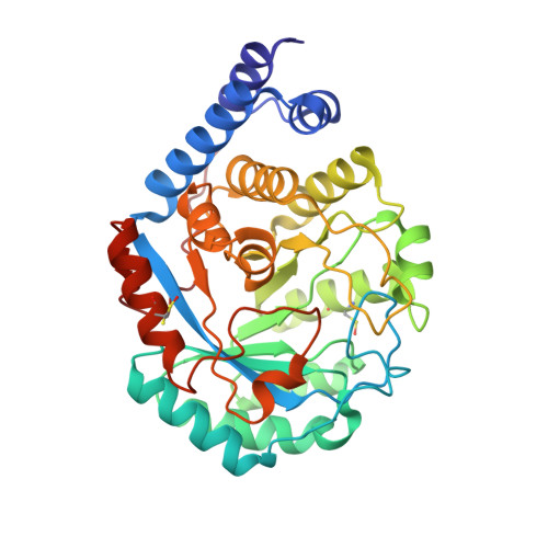
X-ray Structure of the [FeFe]-Hydrogenase Maturase HydE from Thermotoga maritima

Nicolet, Y.Rubach, J.K.Posewitz, M.C.Amara, P.Mathevon, C.Atta, M.Fontecave, M.Fontecilla-Camps, J.C.

(2008) J Biol Chem 283: 18861-18872

  • DOI: https://doi.org/10.1074/jbc.M801161200
  • Primary Citation of Related Structures:  
    3CIW, 3CIX

  • PubMed Abstract: 

    Maturation of the [FeFe]-hydrogenase active site depends on at least the expression of three gene products called HydE, HydF, and HydG. We have solved the high resolution structure of recombinant, reconstituted S-adenosine-L-methionine-dependent HydE from Thermotoga maritima. Besides the conserved [Fe(4)S(4)] cluster involved in the radical-based reaction, this HydE was reported to have a second [Fe(4)S(4)] cluster coordinated by three Cys residues. However, in our crystals, depending on the reconstitution and soaking conditions, this second cluster is either a [Fe(2)S(2)] center, with water occupying the fourth ligand site or is absent. We have carried out site-directed mutagenesis studies on the related HydE from Clostridium acetobutylicum, along with in silico docking and crystal soaking experiments, to define the active site region and three anion-binding sites inside a large, positive cavity, one of which binds SCN(-) with high affinity. Although the overall triose-phosphate isomerase-barrel structure of HydE is very similar to that of biotin synthase, the residues that line the internal cavity are significantly different in the two enzymes.


  • Organizational Affiliation

    Laboratoire de Cristallographie et Cristallogenèse des Protéines, Institut de Biologie Structurale J.P. Ebel, Commissariat à l'Energie Atomique, CNRS, Université J. Fourier, 41 Rue J. Horowitz, 38027 Grenoble Cedex 1, France.


Macromolecules
Find similar proteins by:  (by identity cutoff)  |  3D Structure
Entity ID: 1
MoleculeChains Sequence LengthOrganismDetailsImage
FeFe-Hydrogenase maturase348Thermotoga maritimaMutation(s): 0 
Gene Names: HydE
UniProt
Find proteins for Q9X0Z6 (Thermotoga maritima (strain ATCC 43589 / DSM 3109 / JCM 10099 / NBRC 100826 / MSB8))
Explore Q9X0Z6 
Go to UniProtKB:  Q9X0Z6
Entity Groups  
Sequence Clusters30% Identity50% Identity70% Identity90% Identity95% Identity100% Identity
UniProt GroupQ9X0Z6
Sequence Annotations
Expand
  • Reference Sequence
Small Molecules
Ligands 5 Unique
IDChains Name / Formula / InChI Key2D Diagram3D Interactions
CPS
Query on CPS

Download Ideal Coordinates CCD File 
G [auth A],
I [auth A],
J [auth A],
K [auth A],
L [auth A]
3-[(3-CHOLAMIDOPROPYL)DIMETHYLAMMONIO]-1-PROPANESULFONATE
C32 H58 N2 O7 S
UMCMPZBLKLEWAF-BCTGSCMUSA-N
SAH
Query on SAH

Download Ideal Coordinates CCD File 
F [auth A]S-ADENOSYL-L-HOMOCYSTEINE
C14 H20 N6 O5 S
ZJUKTBDSGOFHSH-WFMPWKQPSA-N
SF4
Query on SF4

Download Ideal Coordinates CCD File 
E [auth A]IRON/SULFUR CLUSTER
Fe4 S4
LJBDFODJNLIPKO-UHFFFAOYSA-N
SAT
Query on SAT

Download Ideal Coordinates CCD File 
H [auth A]sulfoacetic acid
C2 H4 O5 S
AGGIJOLULBJGTQ-UHFFFAOYSA-N
CL
Query on CL

Download Ideal Coordinates CCD File 
B [auth A],
C [auth A],
D [auth A]
CHLORIDE ION
Cl
VEXZGXHMUGYJMC-UHFFFAOYSA-M
Modified Residues  1 Unique
IDChains TypeFormula2D DiagramParent
CSO
Query on CSO
A
L-PEPTIDE LINKINGC3 H7 N O3 SCYS
Experimental Data & Validation

Experimental Data

  • Method: X-RAY DIFFRACTION
  • Resolution: 1.35 Å
  • R-Value Free: 0.177 
  • R-Value Work: 0.143 
  • R-Value Observed: 0.145 
  • Space Group: P 21 21 21
Unit Cell:
Length ( Å )Angle ( ˚ )
a = 50.974α = 90
b = 79.228β = 90
c = 85.941γ = 90
Software Package:
Software NamePurpose
REFMACrefinement
ADSCdata collection
XDSdata reduction
XSCALEdata scaling
SHARPphasing

Structure Validation

View Full Validation Report


Ligand Structure Quality Assessment 


Entry History 

Deposition Data

Revision History  (Full details and data files)

  • Version 1.0: 2008-04-08
    Type: Initial release
  • Version 1.1: 2011-07-13
    Changes: Version format compliance