3CG8

Laccase from Streptomyces coelicolor


Experimental Data Snapshot

  • Method: X-RAY DIFFRACTION
  • Resolution: 2.68 Å
  • R-Value Free: 0.194 
  • R-Value Work: 0.172 
  • R-Value Observed: 0.172 

wwPDB Validation   3D Report Full Report


This is version 1.3 of the entry. See complete history


Literature

The Structure of the Small Laccase from Streptomyces coelicolor Reveals a Link between Laccases and Nitrite Reductases.

Skalova, T.Dohnalek, J.Ostergaard, L.H.Ostergaard, P.R.Kolenko, P.Duskova, J.Stepankova, A.Hasek, J.

(2009) J Mol Biol 385: 1165-1178

  • DOI: https://doi.org/10.1016/j.jmb.2008.11.024
  • Primary Citation of Related Structures:  
    3CG8

  • PubMed Abstract: 

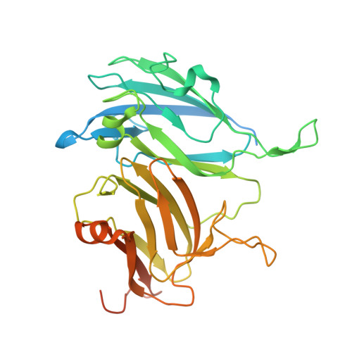
    The X-ray structure of the two-domain laccase (small laccase) from Streptomyces coelicolor A3(2) was solved at 2.7-A resolution. The enzyme differs significantly from all laccases studied structurally so far. It consists of two domains and forms trimers and hence resembles the quaternary structure of nitrite reductases or ceruloplasmins more than that of large laccases. There are three trinuclear copper clusters in the enzyme localized between domains 1 and 2 of each pair of neighbor chains. In this way, a similar geometry of the active site as seen in large laccases is ensured, albeit by different arrangements of domains and protein chains. Three copper ions of type 1 lie close to one another near the surface of the central part of the trimer, and, effectively, a trimeric substrate binding site is formed in their vicinity.


  • Organizational Affiliation

    Institute of Macromolecular Chemistry, Academy of Sciences of the Czech Republic, v.v.i., Heyrovského nám. 2, 162 06 Praha 6, Czech Republic. skalova@imc.cas.cz


Macromolecules
Find similar proteins by:  (by identity cutoff)  |  3D Structure
Entity ID: 1
MoleculeChains Sequence LengthOrganismDetailsImage
laccase
A, B, C
343Streptomyces coelicolorMutation(s): 0 
Gene Names: SC4C6.22
EC: 1.10.3.2
UniProt
Find proteins for Q9XAL8 (Streptomyces coelicolor (strain ATCC BAA-471 / A3(2) / M145))
Explore Q9XAL8 
Go to UniProtKB:  Q9XAL8
Entity Groups  
Sequence Clusters30% Identity50% Identity70% Identity90% Identity95% Identity100% Identity
UniProt GroupQ9XAL8
Sequence Annotations
Expand
  • Reference Sequence
Small Molecules
Ligands 3 Unique
IDChains Name / Formula / InChI Key2D Diagram3D Interactions
PG4
Query on PG4

Download Ideal Coordinates CCD File 
I [auth A],
O [auth B],
P [auth B],
V [auth C],
W [auth C]
TETRAETHYLENE GLYCOL
C8 H18 O5
UWHCKJMYHZGTIT-UHFFFAOYSA-N
CU
Query on CU

Download Ideal Coordinates CCD File 
D [auth A]
E [auth A]
F [auth A]
G [auth A]
J [auth B]
D [auth A],
E [auth A],
F [auth A],
G [auth A],
J [auth B],
K [auth B],
L [auth B],
M [auth B],
Q [auth C],
R [auth C],
S [auth C],
T [auth C]
COPPER (II) ION
Cu
JPVYNHNXODAKFH-UHFFFAOYSA-N
O
Query on O

Download Ideal Coordinates CCD File 
H [auth A],
N [auth B],
U [auth C]
OXYGEN ATOM
O
XLYOFNOQVPJJNP-UHFFFAOYSA-N
Experimental Data & Validation

Experimental Data

Unit Cell:
Length ( Å )Angle ( ˚ )
a = 180.868α = 90
b = 180.868β = 90
c = 177.111γ = 90
Software Package:
Software NamePurpose
DENZOdata reduction
SCALEPACKdata scaling
REFMACrefinement
PDB_EXTRACTdata extraction
MxCuBEdata collection
HKL-2000data reduction
HKL-2000data scaling
SHELXphasing
SHELXDphasing
SHELXEmodel building

Structure Validation

View Full Validation Report


Entry History 

Deposition Data

Revision History  (Full details and data files)

  • Version 1.0: 2009-01-06
    Type: Initial release
  • Version 1.1: 2011-07-13
    Changes: Version format compliance
  • Version 1.2: 2017-10-25
    Changes: Refinement description
  • Version 1.3: 2024-02-21
    Changes: Data collection, Database references, Derived calculations