3CEI

Crystal Structure of Superoxide Dismutase from Helicobacter pylori


Experimental Data Snapshot

  • Method: X-RAY DIFFRACTION
  • Resolution: 2.40 Å
  • R-Value Free: 0.254 
  • R-Value Work: 0.201 

wwPDB Validation   3D Report Full Report


This is version 1.2 of the entry. See complete history


Literature

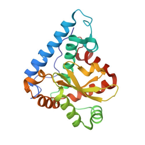
The crystal structure of the superoxide dismutase from Helicobacter pylori reveals a structured C-terminal extension

Esposito, L.Seydel, A.Aiello, R.Sorrentino, G.Cendron, L.Zanotti, G.Zagari, A.

(2008) Biochim Biophys Acta 1784: 1601-1606

  • DOI: https://doi.org/10.1016/j.bbapap.2008.04.024
  • Primary Citation of Related Structures:  
    3CEI

  • PubMed Abstract: 

    Superoxide dismutases (SODs) are key enzymes for fighting oxidative stress. Helicobacter pylori produces a single SOD (HpSOD) which contains iron. The structure of this antioxidant protein has been determined at 2.4 A resolution. It is a dimer of two identical subunits with one iron ion per monomer. The protein shares 53% sequence identity with the corresponding enzyme from Escherichia coli. The model is compared with those of other dimeric Fe-containing SODs. HpSOD shows significant differences in relation to other SODs, the most important being an extended C-terminal tail. This structure provides a model for closely related sequences from species such as Campylobacter, where no structures are currently known. The structure of extended carboxyl termini is discussed in light of putative functions it may serve.


  • Organizational Affiliation

    Institute of Biostructures and Bioimaging, CNR, Via Mezzocannone 16, I-80134, Naples, Italy. luciana.esposito@unina.it


Macromolecules
Find similar proteins by:  (by identity cutoff)  |  3D Structure
Entity ID: 1
MoleculeChains Sequence LengthOrganismDetailsImage
Superoxide dismutase
A, B
213Helicobacter pyloriMutation(s): 0 
Gene Names: sod
EC: 1.15.1.1
UniProt
Find proteins for P43312 (Helicobacter pylori (strain ATCC 700392 / 26695))
Explore P43312 
Go to UniProtKB:  P43312
Entity Groups  
Sequence Clusters30% Identity50% Identity70% Identity90% Identity95% Identity100% Identity
UniProt GroupP43312
Sequence Annotations
Expand
  • Reference Sequence
Experimental Data & Validation

Experimental Data

  • Method: X-RAY DIFFRACTION
  • Resolution: 2.40 Å
  • R-Value Free: 0.254 
  • R-Value Work: 0.201 
  • Space Group: P 63 2 2
Unit Cell:
Length ( Å )Angle ( ˚ )
a = 108.716α = 90
b = 108.716β = 90
c = 158.031γ = 120
Software Package:
Software NamePurpose
CNSrefinement
MOSFLMdata reduction
SCALAdata scaling
AMoREphasing

Structure Validation

View Full Validation Report


Entry History 

Deposition Data

Revision History  (Full details and data files)

  • Version 1.0: 2008-06-10
    Type: Initial release
  • Version 1.1: 2011-07-13
    Changes: Version format compliance
  • Version 1.2: 2023-11-01
    Changes: Data collection, Database references, Derived calculations, Refinement description