3CEH

Human liver glycogen phosphorylase (tense state) in complex with the allosteric inhibitor AVE5688


Experimental Data Snapshot

  • Method: X-RAY DIFFRACTION
  • Resolution: 2.80 Å
  • R-Value Free: 0.247 
  • R-Value Work: 0.165 
  • R-Value Observed: 0.170 

wwPDB Validation   3D Report Full Report


Ligand Structure Quality Assessment 


This is version 1.2 of the entry. See complete history


Literature

Thermodynamic characterization of allosteric glycogen phosphorylase inhibitors.

Anderka, O.Loenze, P.Klabunde, T.Dreyer, M.K.Defossa, E.Wendt, K.U.Schmoll, D.

(2008) Biochemistry 47: 4683-4691

  • DOI: https://doi.org/10.1021/bi702397d
  • Primary Citation of Related Structures:  
    3CEH, 3CEJ, 3CEM

  • PubMed Abstract: 

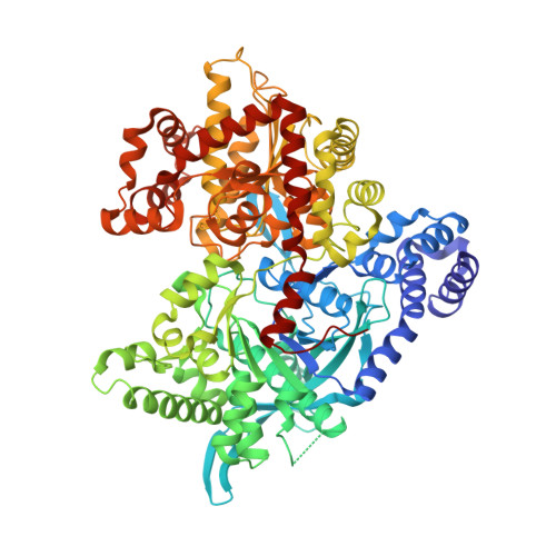
    Glycogen phosphorylase (GP) is a validated target for the treatment of type 2 diabetes. Here we describe highly potent GP inhibitors, AVE5688, AVE2865, and AVE9423. The first two compounds are optimized members of the acyl urea series. The latter represents a novel quinolone class of GP inhibitors, which is introduced in this study. In the enzyme assay, both inhibitor types compete with the physiological activator AMP and act synergistically with glucose. Isothermal titration calorimetry (ITC) shows that the compounds strongly bind to nonphosphorylated, inactive GP (GPb). Binding to phosphorylated, active GP (GPa) is substantially weaker, and the thermodynamic profile reflects a coupled transition to the inactive (tense) conformation. Crystal structures confirm that the three inhibitors bind to the AMP site of tense state GP. These data provide the first direct evidence that acyl urea and quinolone compounds are allosteric inhibitors that selectively bind to and stabilize the inactive conformation of the enzyme. Furthermore, ITC reveals markedly different thermodynamic contributions to inhibitor potency that can be related to the binding modes observed in the cocrystal structures. For AVE5688, which occupies only the lower part of the bifurcated AMP site, binding to GPb (Kd = 170 nM) is exclusively enthalpic (Delta H = -9.0 kcal/mol, TDelta S = 0.3 kcal/mol). The inhibitors AVE2865 (Kd = 9 nM, Delta H = -6.8 kcal/mol, TDelta S = 4.2 kcal/mol) and AVE9423 (Kd = 24 nM, Delta H = -5.9 kcal/mol, TDelta S = 4.6 kcal/mol) fully exploit the volume of the binding pocket. Their pronounced binding entropy can be attributed to the extensive displacement of solvent molecules as well as to ionic interactions with the phosphate recognition site.


  • Organizational Affiliation

    Research and Development, Sanofi Aventis Deutschland GmbH, Frankfurt am Main, Germany. oliver.anderka@sanofi-aventis.com


Macromolecules
Find similar proteins by:  (by identity cutoff)  |  3D Structure
Entity ID: 1
MoleculeChains Sequence LengthOrganismDetailsImage
Glycogen phosphorylase, liver form
A, B
809Homo sapiensMutation(s): 0 
Gene Names: PYGL
EC: 2.4.1.1
UniProt & NIH Common Fund Data Resources
Find proteins for P06737 (Homo sapiens)
Explore P06737 
Go to UniProtKB:  P06737
PHAROS:  P06737
GTEx:  ENSG00000100504 
Entity Groups  
Sequence Clusters30% Identity50% Identity70% Identity90% Identity95% Identity100% Identity
UniProt GroupP06737
Sequence Annotations
Expand
  • Reference Sequence
Small Molecules
Ligands 5 Unique
IDChains Name / Formula / InChI Key2D Diagram3D Interactions
AVE
Query on AVE

Download Ideal Coordinates CCD File 
F [auth A],
K [auth B]
4-[3-(2-Chloro-4,5-difluoro-benzoyl)ureido]-3-trifluoromethoxybenzoic acid
C16 H8 Cl F5 N2 O5
NWQGDIBCFLDHDO-UHFFFAOYSA-N
PLP
Query on PLP

Download Ideal Coordinates CCD File 
E [auth A],
J [auth B]
PYRIDOXAL-5'-PHOSPHATE
C8 H10 N O6 P
NGVDGCNFYWLIFO-UHFFFAOYSA-N
NBG
Query on NBG

Download Ideal Coordinates CCD File 
C [auth A],
G [auth B]
N-acetyl-beta-D-glucopyranosylamine
C8 H15 N O6
IBONACLSSOLHFU-JAJWTYFOSA-N
MES
Query on MES

Download Ideal Coordinates CCD File 
I [auth B]2-(N-MORPHOLINO)-ETHANESULFONIC ACID
C6 H13 N O4 S
SXGZJKUKBWWHRA-UHFFFAOYSA-N
PO4
Query on PO4

Download Ideal Coordinates CCD File 
D [auth A],
H [auth B]
PHOSPHATE ION
O4 P
NBIIXXVUZAFLBC-UHFFFAOYSA-K
Experimental Data & Validation

Experimental Data

  • Method: X-RAY DIFFRACTION
  • Resolution: 2.80 Å
  • R-Value Free: 0.247 
  • R-Value Work: 0.165 
  • R-Value Observed: 0.170 
  • Space Group: P 31
Unit Cell:
Length ( Å )Angle ( ˚ )
a = 124.115α = 90
b = 124.115β = 90
c = 122.491γ = 120
Software Package:
Software NamePurpose
REFMACrefinement
PDB_EXTRACTdata extraction
XDSdata reduction
XSCALEdata scaling

Structure Validation

View Full Validation Report


Ligand Structure Quality Assessment 


Entry History 

Deposition Data

Revision History  (Full details and data files)

  • Version 1.0: 2008-05-27
    Type: Initial release
  • Version 1.1: 2011-07-13
    Changes: Non-polymer description, Version format compliance
  • Version 1.2: 2020-07-29
    Type: Remediation
    Reason: Carbohydrate remediation
    Changes: Data collection, Derived calculations, Structure summary