3C2O

Crystal structure of the quinolinate phosphoribosyl transferase (BNA6) from Sachharomyces cerevisiae complexed with quinolinate


Experimental Data Snapshot

  • Method: X-RAY DIFFRACTION
  • Resolution: 2.30 Å
  • R-Value Free: 0.237 
  • R-Value Work: 0.206 
  • R-Value Observed: 0.207 

wwPDB Validation   3D Report Full Report


Ligand Structure Quality Assessment 


This is version 1.3 of the entry. See complete history


Literature

Comprehensive X-ray Structural Studies of the Quinolinate Phosphoribosyl Transferase (BNA6) from Saccharomyces cerevisiae.

di Luccio, E.Wilson, D.K.

(2008) Biochemistry 47: 4039-4050

  • DOI: https://doi.org/10.1021/bi7020475
  • Primary Citation of Related Structures:  
    3C2E, 3C2O, 3C2R

  • PubMed Abstract: 

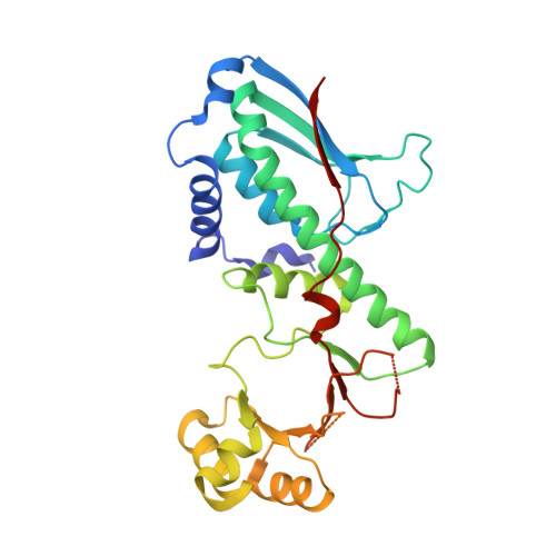
    Quinolinic acid phosphoribosyl transferase (QAPRTase, EC 2.4.2.19) is a 32 kDa enzyme encoded by the BNA6 gene in yeast and catalyzes the formation of nicotinate mononucleotide from quinolinate and 5-phosphoribosyl-1-pyrophosphate (PRPP). QAPRTase plays a key role in the tryptophan degradation pathway via kynurenine, leading to the de novo biosynthesis of NAD (+) and clearing the neurotoxin quinolinate. To improve our understanding of the specificity of the eukaryotic enzyme and the course of events associated with catalysis, we have determined the crystal structures of the apo and singly bound forms with the substrates quinolinate and PRPP. This reveals that the enzyme folds in a manner similar to that of various prokaryotic forms which are approximately 30% identical in sequence. In addition, the structure of the Michaelis complex is approximated by PRPP and the quinolinate analogue phthalate bound to the active site. These results allow insight into the kinetic mechanism of QAPRTase and provide an understanding of structural diversity in the active site of the Saccharomyces cerevisiae enzyme when compared to prokaryotic homologues.


  • Organizational Affiliation

    Section of Molecular and Cellular Biology, University of California, Davis, California 95616, USA.


Macromolecules
Find similar proteins by:  (by identity cutoff)  |  3D Structure
Entity ID: 1
MoleculeChains Sequence LengthOrganismDetailsImage
Nicotinate-nucleotide pyrophosphorylase294Saccharomyces cerevisiaeMutation(s): 0 
Gene Names: BNA6QPT1
EC: 2.4.2.19
UniProt
Find proteins for P43619 (Saccharomyces cerevisiae (strain ATCC 204508 / S288c))
Explore P43619 
Go to UniProtKB:  P43619
Entity Groups  
Sequence Clusters30% Identity50% Identity70% Identity90% Identity95% Identity100% Identity
UniProt GroupP43619
Sequence Annotations
Expand
  • Reference Sequence
Small Molecules
Ligands 1 Unique
IDChains Name / Formula / InChI Key2D Diagram3D Interactions
NTM
Query on NTM

Download Ideal Coordinates CCD File 
B [auth A]QUINOLINIC ACID
C7 H5 N O4
GJAWHXHKYYXBSV-UHFFFAOYSA-N
Binding Affinity Annotations 
IDSourceBinding Affinity
NTM PDBBind:  3C2O Kd: 4.50e+5 (nM) from 1 assay(s)
Binding MOAD:  3C2O Kd: 4.50e+5 (nM) from 1 assay(s)
Experimental Data & Validation

Experimental Data

  • Method: X-RAY DIFFRACTION
  • Resolution: 2.30 Å
  • R-Value Free: 0.237 
  • R-Value Work: 0.206 
  • R-Value Observed: 0.207 
  • Space Group: H 3 2
Unit Cell:
Length ( Å )Angle ( ˚ )
a = 154.802α = 90
b = 154.802β = 90
c = 68.621γ = 120
Software Package:
Software NamePurpose
DENZOdata reduction
SCALEPACKdata scaling
PHASERphasing
REFMACrefinement
PDB_EXTRACTdata extraction
Blu-Icedata collection

Structure Validation

View Full Validation Report


Ligand Structure Quality Assessment 


Entry History 

Deposition Data

Revision History  (Full details and data files)

  • Version 1.0: 2008-03-25
    Type: Initial release
  • Version 1.1: 2011-07-13
    Changes: Version format compliance
  • Version 1.2: 2017-10-25
    Changes: Advisory, Refinement description
  • Version 1.3: 2024-02-21
    Changes: Advisory, Data collection, Database references, Derived calculations