3BOW

Structure of M-calpain in complex with Calpastatin


Experimental Data Snapshot

  • Method: X-RAY DIFFRACTION
  • Resolution: 2.40 Å
  • R-Value Free: 0.258 
  • R-Value Work: 0.198 
  • R-Value Observed: 0.201 

wwPDB Validation   3D Report Full Report


This is version 1.3 of the entry. See complete history


Literature

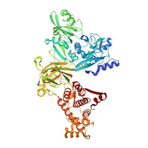
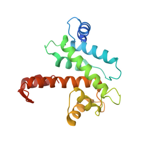
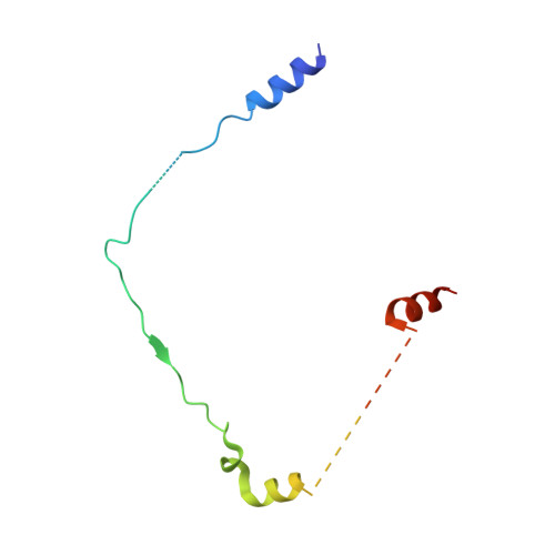
Calcium-bound structure of calpain and its mechanism of inhibition by calpastatin.

Hanna, R.A.Campbell, R.L.Davies, P.L.

(2008) Nature 456: 409-412

  • DOI: https://doi.org/10.1038/nature07451
  • Primary Citation of Related Structures:  
    3BOW

  • PubMed Abstract: 

    Calpains are non-lysosomal calcium-dependent cysteine proteinases that selectively cleave proteins in response to calcium signals and thereby control cellular functions such as cytoskeletal remodelling, cell cycle progression, gene expression and apoptotic cell death. In mammals, the two best-characterized members of the calpain family, calpain 1 and calpain 2 (micro-calpain and m-calpain, respectively), are ubiquitously expressed. The activity of calpains is tightly controlled by the endogenous inhibitor calpastatin, which is an intrinsically unstructured protein capable of reversibly binding and inhibiting four molecules of calpain, but only in the presence of calcium. To date, the mechanism of inhibition by calpastatin and the basis for its absolute specificity have remained speculative. It was not clear how this unstructured protein inhibits calpains without being cleaved itself, nor was it known how calcium induced changes that facilitated the binding of calpastatin to calpain. Here we report the 2.4-A-resolution crystal structure of the calcium-bound calpain 2 heterodimer bound by one of the four inhibitory domains of calpastatin. Calpastatin is seen to inhibit calpain by occupying both sides of the active site cleft. Although the inhibitor passes through the active site cleft it escapes cleavage in a novel manner by looping out and around the active site cysteine. The inhibitory domain of calpastatin recognizes multiple lower affinity sites present only in the calcium-bound form of the enzyme, resulting in an interaction that is tight, specific and calcium dependent. This crystal structure, and that of a related complex, also reveal the conformational changes that calpain undergoes on binding calcium, which include opening of the active site cleft and movement of the domains relative to each other to produce a more compact enzyme.


  • Organizational Affiliation

    Department of Biochemistry, Queen's University, Kingston, Ontario, Canada K7L 3N6.


Macromolecules
Find similar proteins by:  (by identity cutoff)  |  3D Structure
Entity ID: 1
MoleculeChains Sequence LengthOrganismDetailsImage
Calpain-2 catalytic subunit714Rattus norvegicusMutation(s): 1 
Gene Names: Capn2
EC: 3.4.22.53
UniProt
Find proteins for Q07009 (Rattus norvegicus)
Explore Q07009 
Go to UniProtKB:  Q07009
Entity Groups  
Sequence Clusters30% Identity50% Identity70% Identity90% Identity95% Identity100% Identity
UniProt GroupQ07009
Sequence Annotations
Expand
  • Reference Sequence
Find similar proteins by:  (by identity cutoff)  |  3D Structure
Entity ID: 2
MoleculeChains Sequence LengthOrganismDetailsImage
Calpain small subunit 1184Rattus norvegicusMutation(s): 0 
Gene Names: Capns1Capn4Css1
EC: 3.4.22.53
UniProt
Find proteins for Q64537 (Rattus norvegicus)
Explore Q64537 
Go to UniProtKB:  Q64537
Entity Groups  
Sequence Clusters30% Identity50% Identity70% Identity90% Identity95% Identity100% Identity
UniProt GroupQ64537
Sequence Annotations
Expand
  • Reference Sequence
Find similar proteins by:  (by identity cutoff)  |  3D Structure
Entity ID: 3
MoleculeChains Sequence LengthOrganismDetailsImage
Calpastatin95Rattus norvegicusMutation(s): 0 
Gene Names: Cast
UniProt
Find proteins for P27321 (Rattus norvegicus)
Explore P27321 
Go to UniProtKB:  P27321
Entity Groups  
Sequence Clusters30% Identity50% Identity70% Identity90% Identity95% Identity100% Identity
UniProt GroupP27321
Sequence Annotations
Expand
  • Reference Sequence
Experimental Data & Validation

Experimental Data

  • Method: X-RAY DIFFRACTION
  • Resolution: 2.40 Å
  • R-Value Free: 0.258 
  • R-Value Work: 0.198 
  • R-Value Observed: 0.201 
  • Space Group: P 1 21 1
Unit Cell:
Length ( Å )Angle ( ˚ )
a = 67.477α = 90
b = 66.985β = 100.76
c = 108.656γ = 90
Software Package:
Software NamePurpose
MOSFLMdata reduction
SCALAdata scaling
PHASERphasing
REFMACrefinement
PDB_EXTRACTdata extraction

Structure Validation

View Full Validation Report


Entry History 

Deposition Data

Revision History  (Full details and data files)

  • Version 1.0: 2008-11-25
    Type: Initial release
  • Version 1.1: 2011-07-13
    Changes: Advisory, Version format compliance
  • Version 1.2: 2021-10-20
    Changes: Database references, Derived calculations
  • Version 1.3: 2023-08-30
    Changes: Data collection, Refinement description