Crystal Structure of the Major Periplasmic Domain of the Bacterial Membrane Protein Assembly Facilitator YidC.
Oliver, D.C., Paetzel, M.(2008) J Biol Chem 283: 5208-5216
- PubMed: 18093969
- DOI: https://doi.org/10.1074/jbc.M708936200
- Primary Citation of Related Structures:
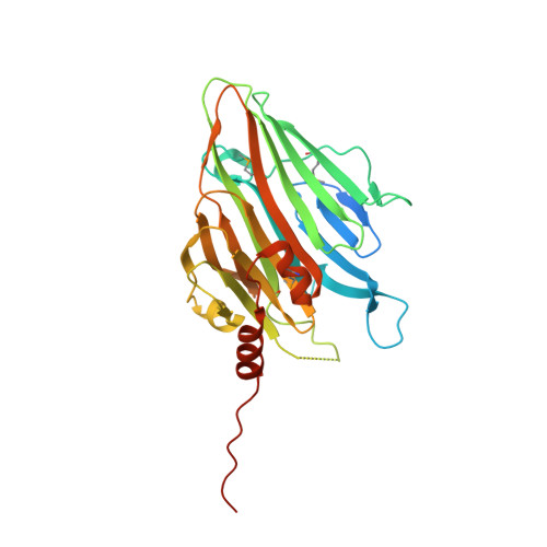
3BLC - PubMed Abstract:
The essential bacterial membrane protein YidC facilitates insertion and assembly of proteins destined for integration into the inner membrane. It has homologues in both mitochondria and chloroplasts. Here we report the crystal structure of the Escherichia coli YidC major periplasmic domain (YidCECP1) at 2.5A resolution. This domain is present in YidC from Gram-negative bacteria and is more than half the size of the full-length protein. The structure reveals that YidCECP1 is made up of a large twisted beta-sandwich protein fold with a C-terminal alpha-helix that packs against one face of the beta-sandwich. Our structure and sequence analysis reveals that the C-terminal alpha-helix and the beta-sheet that it lays against are the most conserved regions of the domain. The region corresponding to the C-terminal alpha-helix was previously shown to be important for the protein insertase function of YidC and is conserved in other YidC-like proteins. The structure reveals that a region of YidC that was previously shown to be involved in binding to SecF maps to one edge of the beta-sandwich. Electrostatic analysis of the molecular surface for this region of YidC reveals a predominantly charged surface and suggests that the SecF-YidC interaction may be electrostatic in nature. Interestingly, YidCECP1 has significant structural similarity to galactose mutarotase from Lactococcus lactis, suggesting that this domain may have another function besides its role in membrane protein assembly.
Organizational Affiliation:
Department of Molecular Biology and Biochemistry, Simon Fraser University, Burnaby, British Columbia V5A 1S6, Canada.