3BC3

Exploring inhibitor binding at the S subsites of cathepsin L


Experimental Data Snapshot

  • Method: X-RAY DIFFRACTION
  • Resolution: 2.20 Å
  • R-Value Free: 0.234 
  • R-Value Work: 0.183 

wwPDB Validation   3D Report Full Report


Ligand Structure Quality Assessment 


This is version 1.4 of the entry. See complete history


Literature

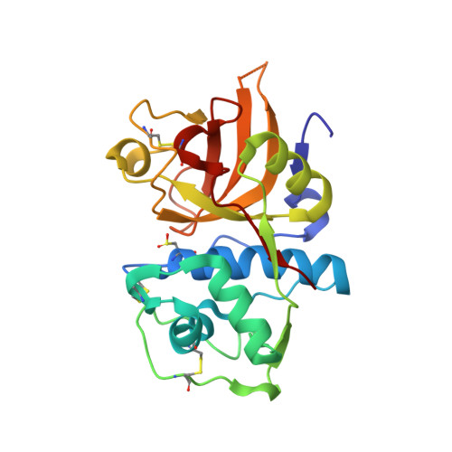
Exploring inhibitor binding at the S' subsites of cathepsin L

Chowdhury, S.F.Joseph, L.Kumar, S.Tulsidas, S.R.Bhat, S.Ziomek, E.Menard, R.Sivaraman, J.Purisima, E.O.

(2008) J Med Chem 51: 1361-1368

  • DOI: https://doi.org/10.1021/jm701190v
  • Primary Citation of Related Structures:  
    3BC3

  • PubMed Abstract: 

    We report a series of noncovalent, reversible inhibitors of cathepsin L that have been designed to explore additional binding interactions with the S' subsites. The design was based on our previously reported crystal structure that suggested the possibility of engineering increased interactions with the S' subsites ( Chowdhury et al. J. Med. Chem. 2002, 45, 5321-5329 ). A representative of these new inhibitors has been co-crystallized with mature cathepsin L, and the structure has been solved and refined at 2.2 A. The inhibitors described in this work extend farther into the S' subsites of cathepsins than any inhibitors reported in the literature thus far. These interactions appear to make use of a S3' subsite that can potentially be exploited for enhanced specificity and/or affinity.


  • Organizational Affiliation

    Biotechnology Research Institute, National Research Council, Canada.


Macromolecules
Find similar proteins by:  (by identity cutoff)  |  3D Structure
Entity ID: 1
MoleculeChains Sequence LengthOrganismDetailsImage
Cathepsin L heavy and light chains
A, B
220Homo sapiensMutation(s): 0 
Gene Names: CTSL
EC: 3.4.22.15
UniProt & NIH Common Fund Data Resources
Find proteins for P07711 (Homo sapiens)
Explore P07711 
Go to UniProtKB:  P07711
PHAROS:  P07711
GTEx:  ENSG00000135047 
Entity Groups  
Sequence Clusters30% Identity50% Identity70% Identity90% Identity95% Identity100% Identity
UniProt GroupP07711
Sequence Annotations
Expand
  • Reference Sequence
Small Molecules
Ligands 1 Unique
IDChains Name / Formula / InChI Key2D Diagram3D Interactions
OPT
Query on OPT

Download Ideal Coordinates CCD File 
C [auth A],
D [auth B]
S-benzyl-N-(biphenyl-4-ylacetyl)-L-cysteinyl-N~5~-(diaminomethyl)-D-ornithyl-N-(2-phenylethyl)-L-tyrosinamide
C47 H55 N7 O5 S
IMZNRHOMBTZKJS-IWSHAHEXSA-N
Modified Residues  1 Unique
IDChains TypeFormula2D DiagramParent
CSD
Query on CSD
A, B
L-PEPTIDE LINKINGC3 H7 N O4 SCYS
Binding Affinity Annotations 
IDSourceBinding Affinity
OPT PDBBind:  3BC3 Ki: 155 (nM) from 1 assay(s)
Binding MOAD:  3BC3 Ki: 155 (nM) from 1 assay(s)
Experimental Data & Validation

Experimental Data

  • Method: X-RAY DIFFRACTION
  • Resolution: 2.20 Å
  • R-Value Free: 0.234 
  • R-Value Work: 0.183 
  • Space Group: C 2 2 21
Unit Cell:
Length ( Å )Angle ( ˚ )
a = 74.765α = 90
b = 90.929β = 90
c = 126.026γ = 90
Software Package:
Software NamePurpose
MOLREPphasing
CNSrefinement
PDB_EXTRACTdata extraction
CrystalCleardata collection
DENZOdata reduction
SCALEPACKdata scaling

Structure Validation

View Full Validation Report


Ligand Structure Quality Assessment 


Entry History 

Deposition Data

Revision History  (Full details and data files)

  • Version 1.0: 2008-03-18
    Type: Initial release
  • Version 1.1: 2011-07-13
    Changes: Version format compliance
  • Version 1.2: 2014-11-12
    Changes: Structure summary
  • Version 1.3: 2017-10-25
    Changes: Refinement description
  • Version 1.4: 2023-08-30
    Changes: Data collection, Database references, Derived calculations, Refinement description