3B9R

SERCA Ca2+-ATPase E2 aluminium fluoride complex without thapsigargin


Experimental Data Snapshot

  • Method: X-RAY DIFFRACTION
  • Resolution: 3.00 Å
  • R-Value Free: 0.235 
  • R-Value Work: 0.185 
  • R-Value Observed: 0.186 

wwPDB Validation   3D Report Full Report


Ligand Structure Quality Assessment 


This is version 1.2 of the entry. See complete history


Literature

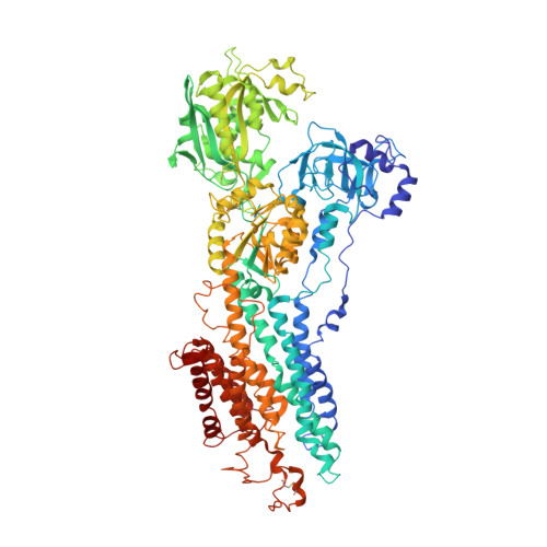
The structural basis of calcium transport by the calcium pump

Olesen, C.Picard, M.Winther, A.M.L.Gyrup, C.Morth, J.P.Oxvig, C.Moller, J.V.Nissen, P.

(2007) Nature 450: 1036-1042

  • DOI: https://doi.org/10.1038/nature06418
  • Primary Citation of Related Structures:  
    3B9B, 3B9R, 3BA6

  • PubMed Abstract: 

    The sarcoplasmic reticulum Ca2+-ATPase, a P-type ATPase, has a critical role in muscle function and metabolism. Here we present functional studies and three new crystal structures of the rabbit skeletal muscle Ca2+-ATPase, representing the phosphoenzyme intermediates associated with Ca2+ binding, Ca2+ translocation and dephosphorylation, that are based on complexes with a functional ATP analogue, beryllium fluoride and aluminium fluoride, respectively. The structures complete the cycle of nucleotide binding and cation transport of Ca2+-ATPase. Phosphorylation of the enzyme triggers the onset of a conformational change that leads to the opening of a luminal exit pathway defined by the transmembrane segments M1 through M6, which represent the canonical membrane domain of P-type pumps. Ca2+ release is promoted by translocation of the M4 helix, exposing Glu 309, Glu 771 and Asn 796 to the lumen. The mechanism explains how P-type ATPases are able to form the steep electrochemical gradients required for key functions in eukaryotic cells.


  • Organizational Affiliation

    Centre for Membrane Pumps in Cells and Disease-PUMPKIN, Danish National Research Foundation, University of Aarhus, Ole Worms Alle, blg. 1185, DK - 8000 Aarhus C, Denmark.


Macromolecules
Find similar proteins by:  (by identity cutoff)  |  3D Structure
Entity ID: 1
MoleculeChains Sequence LengthOrganismDetailsImage
Sarcoplasmic/endoplasmic reticulum calcium ATPase 1
A, B
994Oryctolagus cuniculusMutation(s): 0 
EC: 3.6.3.8
Membrane Entity: Yes 
UniProt
Find proteins for P04191 (Oryctolagus cuniculus)
Explore P04191 
Go to UniProtKB:  P04191
Entity Groups  
Sequence Clusters30% Identity50% Identity70% Identity90% Identity95% Identity100% Identity
UniProt GroupP04191
Sequence Annotations
Expand
  • Reference Sequence
Small Molecules
Ligands 4 Unique
IDChains Name / Formula / InChI Key2D Diagram3D Interactions
ACP
Query on ACP

Download Ideal Coordinates CCD File 
F [auth A],
J [auth B]
PHOSPHOMETHYLPHOSPHONIC ACID ADENYLATE ESTER
C11 H18 N5 O12 P3
UFZTZBNSLXELAL-IOSLPCCCSA-N
ALF
Query on ALF

Download Ideal Coordinates CCD File 
C [auth A],
G [auth B]
TETRAFLUOROALUMINATE ION
Al F4
UYOMQIYKOOHAMK-UHFFFAOYSA-J
K
Query on K

Download Ideal Coordinates CCD File 
E [auth A],
I [auth B]
POTASSIUM ION
K
NPYPAHLBTDXSSS-UHFFFAOYSA-N
MG
Query on MG

Download Ideal Coordinates CCD File 
D [auth A],
H [auth B]
MAGNESIUM ION
Mg
JLVVSXFLKOJNIY-UHFFFAOYSA-N
Experimental Data & Validation

Experimental Data

  • Method: X-RAY DIFFRACTION
  • Resolution: 3.00 Å
  • R-Value Free: 0.235 
  • R-Value Work: 0.185 
  • R-Value Observed: 0.186 
  • Space Group: P 1 21 1
Unit Cell:
Length ( Å )Angle ( ˚ )
a = 131.98α = 90
b = 94.43β = 107.79
c = 136.18γ = 90
Software Package:
Software NamePurpose
XSCALEdata scaling
DMphasing
PHENIXrefinement
PDB_EXTRACTdata extraction
ADSCdata collection
XDSdata reduction
PHASERphasing

Structure Validation

View Full Validation Report


Ligand Structure Quality Assessment 


Entry History 

Deposition Data

Revision History  (Full details and data files)

  • Version 1.0: 2007-12-18
    Type: Initial release
  • Version 1.1: 2011-07-13
    Changes: Version format compliance
  • Version 1.2: 2023-11-01
    Changes: Data collection, Database references, Derived calculations, Refinement description