Structures and solution properties of two novel periplasmic sensor domains with c-type heme from chemotaxis proteins of Geobacter sulfurreducens: implications for signal transduction.
Pokkuluri, P.R., Pessanha, M., Londer, Y.Y., Wood, S.J., Duke, N.E., Wilton, R., Catarino, T., Salgueiro, C.A., Schiffer, M.(2008) J Mol Biol 377: 1498-1517
- PubMed: 18329666
- DOI: https://doi.org/10.1016/j.jmb.2008.01.087
- Primary Citation of Related Structures:
3B42, 3B47 - PubMed Abstract:
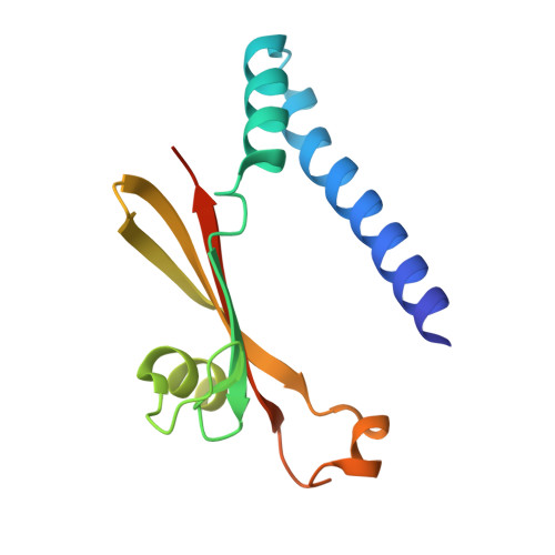
Periplasmic sensor domains from two methyl-accepting chemotaxis proteins from Geobacter sulfurreducens (encoded by genes GSU0935 and GSU0582) were expressed in Escherichia coli. The sensor domains were isolated, purified, characterized in solution, and their crystal structures were determined. In the crystal, both sensor domains form swapped dimers and show a PAS-type fold. The swapped segment consists of two helices of about 45 residues at the N terminus with the hemes located between the two monomers. In the case of the GSU0582 sensor, the dimer contains a crystallographic 2-fold symmetry and the heme is coordinated by an axial His and a water molecule. In the case of the GSU0935 sensor, the crystals contain a non-crystallographic dimer, and surprisingly, the coordination of the heme in each monomer is different; monomer A heme has His-Met ligation and monomer B heme has His-water ligation as found in the GSU0582 sensor. The structures of these sensor domains are the first structures of PAS domains containing covalently bound heme. Optical absorption, electron paramagnetic resonance and NMR spectroscopy have revealed that the heme groups of both sensor domains are high-spin and low-spin in the oxidized and reduced forms, respectively, and that the spin-state interconversion involves a heme axial ligand replacement. Both sensor domains bind NO in their ferric and ferrous forms but bind CO only in the reduced form. The binding of both NO and CO occurs via an axial ligand exchange process, and is fully reversible. The reduction potentials of the sensor domains differ by 95 mV (-156 mV and -251 mV for sensors GSU0582 and GSU0935, respectively). The swapped dimerization of these sensor domains and redox-linked ligand switch might be related to the mechanism of signal transduction by these chemotaxis proteins.
Organizational Affiliation:
Biosciences Division, Argonne National Laboratory, Argonne, IL 60439, USA.