3B2D

Crystal structure of human RP105/MD-1 complex


Experimental Data Snapshot

  • Method: X-RAY DIFFRACTION
  • Resolution: 2.80 Å
  • R-Value Free: 0.284 
  • R-Value Work: 0.218 
  • R-Value Observed: 0.221 

wwPDB Validation   3D Report Full Report


Ligand Structure Quality Assessment 


This is version 2.0 of the entry. See complete history


Literature

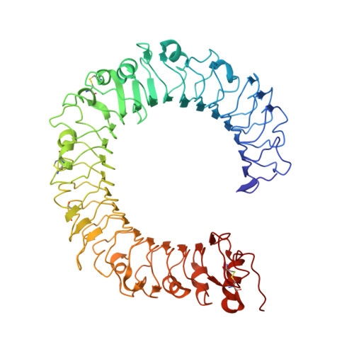
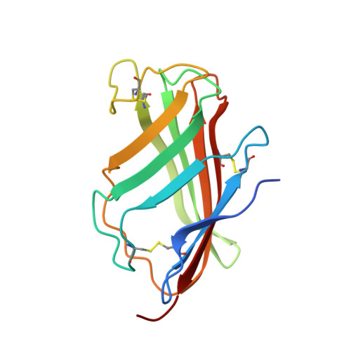
Crystal Structures of Mouse and Human RP105/MD-1 Complexes Reveal Unique Dimer Organization of the Toll-Like Receptor Family.

Ohto, U.Miyake, K.Shimizu, T.

(2011) J Mol Biol 413: 815-825

  • DOI: https://doi.org/10.1016/j.jmb.2011.09.020
  • Primary Citation of Related Structures:  
    3B2D, 3T6Q

  • PubMed Abstract: 

    The Toll-like receptor (TLR) 4/MD-2 heterodimer senses lipopolysaccharide (LPS). RP105 (radioprotective 105 kDa), a TLR-related molecule, is similar to TLR4 in that the extracellular leucine-rich repeats associate with MD-1, the MD-2-like molecule. MD-2 has a unique hydrophobic cavity that directly binds to lipid A, the active center of LPS. LPS-bound MD-2 opens the secondary interface with TLR4, leading to dimerization of TLR4/MD-2. MD-1 also has a hydrophobic cavity that accommodates lipid IVa, a precursor of lipid A, suggesting a role for the RP105/MD-1 heterodimer in sensing LPS or related microbial products. Little is known, however, about the structure of the RP105/MD-1 heterodimer or its oligomer. Here, we have determined the crystal structures of mouse and human RP105/MD-1 complexes at 1.9 and 2.8 Å resolutions, respectively. Both mouse and human RP105/MD-1 exhibit dimerization of the 1:1 RP105/MD-1 complex, demonstrating a novel organization. The "m"-shaped 2:2 RP105/MD-1 complex exhibits an inverse arrangement, with N-termini interacting in the middle. Thus, the dimerization interface of RP105/MD-1 is located on the opposite side of the complex, compared to the 2:2 TLR4/MD-2 complex. These results demonstrate that the 2:2 RP105/MD-1 complex is distinct from previously reported TLR dimers, including TLR4/MD-2, TLR1/TLR2, TLR2/TLR6, and TLR3, all of which facilitate homotypic or heterotypic interaction of the C-terminal cytoplasmic signaling domain.


  • Organizational Affiliation

    Graduate School of Pharmaceutical Sciences, University of Tokyo, Hongo, Bunkyo-ku, Tokyo 113-0033, Japan.


Macromolecules
Find similar proteins by:  (by identity cutoff)  |  3D Structure
Entity ID: 1
MoleculeChains Sequence LengthOrganismDetailsImage
CD180 antigen
A, B
603Homo sapiensMutation(s): 0 
Gene Names: CD180LY64RP105
UniProt & NIH Common Fund Data Resources
Find proteins for Q99467 (Homo sapiens)
Explore Q99467 
Go to UniProtKB:  Q99467
PHAROS:  Q99467
GTEx:  ENSG00000134061 
Entity Groups  
Sequence Clusters30% Identity50% Identity70% Identity90% Identity95% Identity100% Identity
UniProt GroupQ99467
Sequence Annotations
Expand
  • Reference Sequence
Find similar proteins by:  (by identity cutoff)  |  3D Structure
Entity ID: 2
MoleculeChains Sequence LengthOrganismDetailsImage
Lymphocyte antigen 86
C, D
144Homo sapiensMutation(s): 0 
Gene Names: LY86MD1
UniProt & NIH Common Fund Data Resources
Find proteins for O95711 (Homo sapiens)
Explore O95711 
Go to UniProtKB:  O95711
PHAROS:  O95711
GTEx:  ENSG00000112799 
Entity Groups  
Sequence Clusters30% Identity50% Identity70% Identity90% Identity95% Identity100% Identity
UniProt GroupO95711
Sequence Annotations
Expand
  • Reference Sequence
Oligosaccharides

Help

Entity ID: 3
MoleculeChains Length2D Diagram Glycosylation3D Interactions
alpha-D-mannopyranose-(1-2)-alpha-D-mannopyranose-(1-3)-[alpha-D-mannopyranose-(1-2)-alpha-D-mannopyranose-(1-6)]alpha-D-mannopyranose-(1-6)-[alpha-D-mannopyranose-(1-3)]beta-D-mannopyranose-(1-4)-2-acetamido-2-deoxy-beta-D-glucopyranose-(1-4)-2-acetamido-2-deoxy-beta-D-glucopyranose
E
9N-Glycosylation
Glycosylation Resources
GlyTouCan:  G37135JQ
GlyCosmos:  G37135JQ
GlyGen:  G37135JQ
Entity ID: 4
MoleculeChains Length2D Diagram Glycosylation3D Interactions
2-acetamido-2-deoxy-beta-D-glucopyranose-(1-4)-2-acetamido-2-deoxy-beta-D-glucopyranose
F, H
2N-Glycosylation
Glycosylation Resources
GlyTouCan:  G42666HT
GlyCosmos:  G42666HT
GlyGen:  G42666HT
Entity ID: 5
MoleculeChains Length2D Diagram Glycosylation3D Interactions
alpha-D-mannopyranose-(1-2)-alpha-D-mannopyranose-(1-6)-[alpha-D-mannopyranose-(1-3)]alpha-D-mannopyranose-(1-6)-[alpha-D-mannopyranose-(1-3)]beta-D-mannopyranose-(1-4)-2-acetamido-2-deoxy-beta-D-glucopyranose-(1-4)-2-acetamido-2-deoxy-beta-D-glucopyranose
G
8N-Glycosylation
Glycosylation Resources
GlyTouCan:  G61846BY
GlyCosmos:  G61846BY
GlyGen:  G61846BY
Small Molecules
Ligands 1 Unique
IDChains Name / Formula / InChI Key2D Diagram3D Interactions
NAG
Query on NAG

Download Ideal Coordinates CCD File 
I [auth B]2-acetamido-2-deoxy-beta-D-glucopyranose
C8 H15 N O6
OVRNDRQMDRJTHS-FMDGEEDCSA-N
Experimental Data & Validation

Experimental Data

  • Method: X-RAY DIFFRACTION
  • Resolution: 2.80 Å
  • R-Value Free: 0.284 
  • R-Value Work: 0.218 
  • R-Value Observed: 0.221 
  • Space Group: P 21 21 2
Unit Cell:
Length ( Å )Angle ( ˚ )
a = 203.997α = 90
b = 66.441β = 90
c = 119.097γ = 90
Software Package:
Software NamePurpose
MOLREPphasing
REFMACrefinement
HKL-2000data reduction
HKL-2000data scaling

Structure Validation

View Full Validation Report


Ligand Structure Quality Assessment 


Entry History 

Deposition Data

Revision History  (Full details and data files)

  • Version 1.0: 2011-11-09
    Type: Initial release
  • Version 2.0: 2020-07-29
    Type: Remediation
    Reason: Carbohydrate remediation
    Changes: Atomic model, Data collection, Database references, Derived calculations, Structure summary