Structures and reaction mechanisms of the two related enzymes, PurN and PurU.
Sampei, G., Kanagawa, M., Baba, S., Shimasaki, T., Taka, H., Mitsui, S., Fujiwara, S., Yanagida, Y., Kusano, M., Suzuki, S., Terao, K., Kawai, H., Fukai, Y., Nakagawa, N., Ebihara, A., Kuramitsu, S., Yokoyama, S., Kawai, G.(2013) J Biochem 154: 569-579
- PubMed: 24108189
- DOI: https://doi.org/10.1093/jb/mvt090
- Primary Citation of Related Structures:
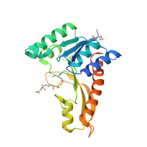
2YWR, 3AUF, 3AV3, 3W7B - PubMed Abstract:
The crystal structures of glycinamide ribonucleotide transformylases (PurNs) from Aquifex aeolicus (Aa), Geobacillus kaustophilus (Gk) and Symbiobacterium toebii (St), and of formyltetrahydrofolate hydrolase (PurU) from Thermus thermophilus (Tt) were determined. The monomer structures of the determined PurN and PurU were very similar to the known structure of PurN, but oligomeric states were different; AaPurN and StPurN formed dimers, GkPurN formed monomer and PurU formed tetramer in the crystals. PurU had a regulatory ACT domain in its N-terminal side. So far several structures of PurUs have been determined, yet, the mechanisms of the catalysis and the regulation of PurU have not been elucidated. We, therefore, modelled ligand-bound structures of PurN and PurU, and performed molecular dynamics simulations to elucidate the reaction mechanisms. The evolutionary relationship of the two enzymes is discussed based on the comparisons of the structures and the catalytic mechanisms.
Organizational Affiliation:
Department of Engineering Science, Graduate School of Informatics and Engineering, The University of Electro-Communications, 1-5-1 Chofugaoka, Chofu, Tokyo 182-8585, Japan; RIKEN SPring-8 Center, Harima Institute, 1-1-1 Kouto, Sayo, Sayo, Hyogo 679-5148, Japan; Structural Biology Group, SPring-8/JASRI, 1-1-1 Kouto, Sayo, Sayo, Hyogo 679-5198, Japan; Department of Life and Environmental Sciences, Faculty of Engineering, Chiba Institute of Technology, 2-17-1 Tsudanuma, Narashino, Chiba 275-0016, Japan; and Department of Biological Sciences, Graduate School of Science, Osaka University, 1-1 Machikaneyama-cho, Toyonaka, Osaka 560-0043, Japan.