3AUJ

Structure of diol dehydratase complexed with glycerol


Experimental Data Snapshot

  • Method: X-RAY DIFFRACTION
  • Resolution: 2.10 Å
  • R-Value Free: 0.191 
  • R-Value Work: 0.186 
  • R-Value Observed: 0.186 

wwPDB Validation   3D Report Full Report


Ligand Structure Quality Assessment 


This is version 1.3 of the entry. See complete history


Literature

Redesign of coenzyme B(12) dependent diol dehydratase to be resistant to the mechanism-based inactivation by glycerol and act on longer chain 1,2-diols

Yamanishi, M.Kinoshita, K.Fukuoka, M.Saito, T.Tanokuchi, A.Ikeda, Y.Obayashi, H.Mori, K.Shibata, N.Tobimatsu, T.Toraya, T.

(2012) FEBS J 279: 793-804

  • DOI: https://doi.org/10.1111/j.1742-4658.2012.08470.x
  • Primary Citation of Related Structures:  
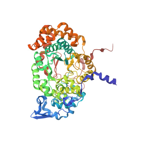
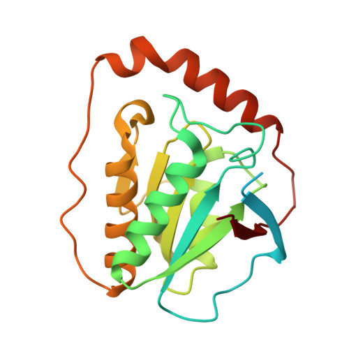
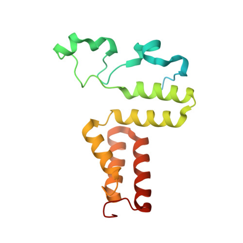
    3AUJ

  • PubMed Abstract: 

    Coenzyme B(12) dependent diol dehydratase undergoes mechanism-based inactivation by glycerol, accompanying the irreversible cleavage of the coenzyme Co-C bond. Bachovchin et al. [Biochemistry16, 1082-1092 (1977)] reported that glycerol bound in the G(S) conformation, in which the pro-S-CH(2) OH group is oriented to the hydrogen-abstracting site, primarily contributes to the inactivation reaction. To understand the mechanism of inactivation by glycerol, we analyzed the X-ray structure of diol dehydratase complexed with cyanocobalamin and glycerol. Glycerol is bound to the active site preferentially in the same conformation as that of (S)-1,2-propanediol, i.e. in the G(S) conformation, with its 3-OH group hydrogen bonded to Serα301, but not to nearby Glnα336. k(inact) of the Sα301A, Qα336A and Sα301A/Qα336A mutants with glycerol was much smaller than that of the wild-type enzyme. k(cat) /k(inact) showed that the Sα301A and Qα336A mutants are substantially more resistant to glycerol inactivation than the wild-type enzyme, suggesting that Serα301 and Glnα336 are directly or indirectly involved in the inactivation. The degree of preference for (S)-1,2-propanediol decreased on these mutations. The substrate activities towards longer chain 1,2-diols significantly increased on the Sα301A/Qα336A double mutation, probably because these amino acid substitutions yield more space for accommodating a longer alkyl group on C3 of 1,2-diols. Database Structural data are available in the Protein Data Bank under the accession number 3AUJ. Structured digital abstract • Diol dehydrase gamma subunit, Diol dehydrase beta subunit and Diol dehydrase alpha subunit physically interact by X-ray crystallography (View interaction).


  • Organizational Affiliation

    Department of Bioscience and Biotechnology, Graduate School of Natural Science and Technology, Okayama University, Japan.


Macromolecules
Find similar proteins by:  (by identity cutoff)  |  3D Structure
Entity ID: 1
MoleculeChains Sequence LengthOrganismDetailsImage
Diol dehydrase alpha subunitA,
D [auth L]
554Klebsiella oxytocaMutation(s): 0 
Gene Names: pddA
EC: 4.2.1.28
UniProt
Find proteins for Q59470 (Klebsiella oxytoca)
Explore Q59470 
Go to UniProtKB:  Q59470
Entity Groups  
Sequence Clusters30% Identity50% Identity70% Identity90% Identity95% Identity100% Identity
UniProt GroupQ59470
Sequence Annotations
Expand
  • Reference Sequence
Find similar proteins by:  (by identity cutoff)  |  3D Structure
Entity ID: 2
MoleculeChains Sequence LengthOrganismDetailsImage
Diol dehydrase beta subunit
B, E
224Klebsiella oxytocaMutation(s): 0 
Gene Names: pddB
EC: 4.2.1.28
UniProt
Find proteins for Q59471 (Klebsiella oxytoca)
Explore Q59471 
Go to UniProtKB:  Q59471
Entity Groups  
Sequence Clusters30% Identity50% Identity70% Identity90% Identity95% Identity100% Identity
UniProt GroupQ59471
Sequence Annotations
Expand
  • Reference Sequence
Find similar proteins by:  (by identity cutoff)  |  3D Structure
Entity ID: 3
MoleculeChains Sequence LengthOrganismDetailsImage
Diol dehydrase gamma subunitC [auth G],
F [auth M]
173Klebsiella oxytocaMutation(s): 0 
Gene Names: pddC
EC: 4.2.1.28
UniProt
Find proteins for Q59472 (Klebsiella oxytoca)
Explore Q59472 
Go to UniProtKB:  Q59472
Entity Groups  
Sequence Clusters30% Identity50% Identity70% Identity90% Identity95% Identity100% Identity
UniProt GroupQ59472
Sequence Annotations
Expand
  • Reference Sequence
Small Molecules
Ligands 4 Unique
IDChains Name / Formula / InChI Key2D Diagram3D Interactions
B12
Query on B12

Download Ideal Coordinates CCD File 
J [auth B],
P [auth E]
COBALAMIN
C62 H89 Co N13 O14 P
LKVIQTCSMMVGFU-DWSMJLPVSA-N
PO4
Query on PO4

Download Ideal Coordinates CCD File 
I [auth A]
K [auth B]
L [auth G]
O [auth L]
Q [auth E]
I [auth A],
K [auth B],
L [auth G],
O [auth L],
Q [auth E],
R [auth M]
PHOSPHATE ION
O4 P
NBIIXXVUZAFLBC-UHFFFAOYSA-K
GOL
Query on GOL

Download Ideal Coordinates CCD File 
G [auth A],
M [auth L]
GLYCEROL
C3 H8 O3
PEDCQBHIVMGVHV-UHFFFAOYSA-N
CA
Query on CA

Download Ideal Coordinates CCD File 
H [auth A],
N [auth L]
CALCIUM ION
Ca
BHPQYMZQTOCNFJ-UHFFFAOYSA-N
Experimental Data & Validation

Experimental Data

  • Method: X-RAY DIFFRACTION
  • Resolution: 2.10 Å
  • R-Value Free: 0.191 
  • R-Value Work: 0.186 
  • R-Value Observed: 0.186 
  • Space Group: C 1 2 1
Unit Cell:
Length ( Å )Angle ( ˚ )
a = 190.61α = 90
b = 196.8β = 95.03
c = 77.36γ = 90
Software Package:
Software NamePurpose
BSSdata collection
CNSrefinement
HKL-2000data reduction
HKL-2000data scaling
CNSphasing

Structure Validation

View Full Validation Report


Ligand Structure Quality Assessment 


Entry History 

Deposition Data

Revision History  (Full details and data files)

  • Version 1.0: 2012-02-22
    Type: Initial release
  • Version 1.1: 2012-10-24
    Changes: Non-polymer description
  • Version 1.2: 2013-06-19
    Changes: Database references
  • Version 1.3: 2023-11-01
    Changes: Data collection, Database references, Derived calculations, Refinement description