3AOT

Crystal structure of juvenile hormone binding protein from silkworm in its apo form


Experimental Data Snapshot

  • Method: X-RAY DIFFRACTION
  • Resolution: 2.20 Å
  • R-Value Free: 0.255 
  • R-Value Work: 0.234 
  • R-Value Observed: 0.235 

wwPDB Validation   3D Report Full Report


This is version 1.3 of the entry. See complete history


Literature

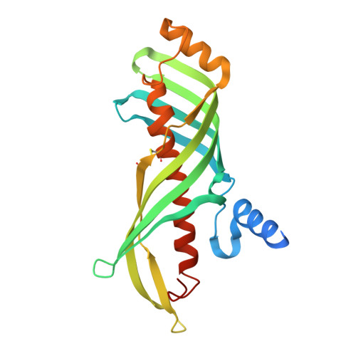
Structural mechanism of JH delivery in hemolymph by JHBP of silkworm, Bombyx mori.

Suzuki, R.Fujimoto, Z.Shiotsuki, T.Tsuchiya, W.Momma, M.Tase, A.Miyazawa, M.Yamazaki, T.

(2011) Sci Rep 1: 133-133

  • DOI: https://doi.org/10.1038/srep00133
  • Primary Citation of Related Structures:  
    2RQF, 3AOS, 3AOT

  • PubMed Abstract: 

    Juvenile hormone (JH) plays crucial roles in many aspects of the insect life. All the JH actions are initiated by transport of JH in the hemolymph as a complex with JH-binding protein (JHBP) to target tissues. Here, we report structural mechanism of JH delivery by JHBP based upon the crystal and solution structures of apo and JH-bound JHBP. In solution, apo-JHBP exists in equilibrium of multiple conformations with different orientations of the gate helix for the hormone-binding pocket ranging from closed to open forms. JH-binding to the gate-open form results in the fully closed JHBP-JH complex structure where the bound JH is completely buried inside the protein. JH-bound JHBP opens the gate helix to release the bound hormone likely by sensing the less polar environment at the membrane surface of target cells. This is the first report that provides structural insight into JH signaling.


  • Organizational Affiliation

    Biomolecular Research Unit, National Institute of Agrobiological Sciences, 2-1-2 Kannondai, Tsukuba, Ibaraki 305-8602, Japan.


Macromolecules
Find similar proteins by:  (by identity cutoff)  |  3D Structure
Entity ID: 1
MoleculeChains Sequence LengthOrganismDetailsImage
Hemolymph juvenile hormone binding protein227Bombyx moriMutation(s): 0 
Gene Names: hJHBP
UniProt
Find proteins for Q9U556 (Bombyx mori)
Explore Q9U556 
Go to UniProtKB:  Q9U556
Entity Groups  
Sequence Clusters30% Identity50% Identity70% Identity90% Identity95% Identity100% Identity
UniProt GroupQ9U556
Sequence Annotations
Expand
  • Reference Sequence
Small Molecules
Ligands 3 Unique
IDChains Name / Formula / InChI Key2D Diagram3D Interactions
PG4
Query on PG4

Download Ideal Coordinates CCD File 
B [auth A],
C [auth A],
D [auth A],
E [auth A],
F [auth A]
TETRAETHYLENE GLYCOL
C8 H18 O5
UWHCKJMYHZGTIT-UHFFFAOYSA-N
PGE
Query on PGE

Download Ideal Coordinates CCD File 
G [auth A]TRIETHYLENE GLYCOL
C6 H14 O4
ZIBGPFATKBEMQZ-UHFFFAOYSA-N
PEG
Query on PEG

Download Ideal Coordinates CCD File 
H [auth A],
I [auth A],
J [auth A]
DI(HYDROXYETHYL)ETHER
C4 H10 O3
MTHSVFCYNBDYFN-UHFFFAOYSA-N
Experimental Data & Validation

Experimental Data

  • Method: X-RAY DIFFRACTION
  • Resolution: 2.20 Å
  • R-Value Free: 0.255 
  • R-Value Work: 0.234 
  • R-Value Observed: 0.235 
  • Space Group: P 63 2 2
Unit Cell:
Length ( Å )Angle ( ˚ )
a = 90.898α = 90
b = 90.898β = 90
c = 138.336γ = 120
Software Package:
Software NamePurpose
HKL-2000data collection
MOLREPphasing
REFMACrefinement
HKL-2000data reduction
SCALEPACKdata scaling

Structure Validation

View Full Validation Report


Entry History 

Deposition Data

Revision History  (Full details and data files)

  • Version 1.0: 2011-10-12
    Type: Initial release
  • Version 1.1: 2012-02-15
    Changes: Database references
  • Version 1.2: 2013-06-19
    Changes: Database references
  • Version 1.3: 2023-11-01
    Changes: Data collection, Database references, Derived calculations, Refinement description