3AJ3

Crystal structure of selenomethionine substituted 4-pyridoxolactonase from Mesorhizobium loti


Experimental Data Snapshot

  • Method: X-RAY DIFFRACTION
  • Resolution: 1.58 Å
  • R-Value Free: 0.190 
  • R-Value Work: 0.163 
  • R-Value Observed: 0.164 

wwPDB Validation   3D Report Full Report


This is version 1.3 of the entry. See complete history


Literature

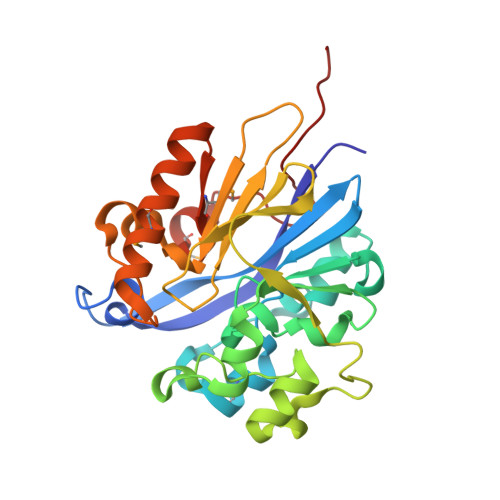
Structure of 4-pyridoxolactonase from Mesorhizobium loti.

Kobayashi, J.Yoshikane, Y.Yagi, T.Baba, S.Mizutani, K.Takahashi, N.Mikami, B.

(2014) Acta Crystallogr F Struct Biol Commun 70: 424-432

  • DOI: https://doi.org/10.1107/S2053230X14003926
  • Primary Citation of Related Structures:  
    3AJ3, 4KEP, 4KEQ

  • PubMed Abstract: 

    4-Pyridoxolactonase from Mesorhizobium loti catalyzes the zinc-dependent lactone-ring hydrolysis of 4-pyridoxolactone (4PAL) to 4-pyridoxic acid (4PA) in vitamin B6 degradation pathway I. The crystal structures of 4-pyridoxolactonase and its complex with 5-pyridoxolactone (5PAL; the competitive inhibitor) were determined. The overall structure was an αβ/βα sandwich fold, and two zinc ions were coordinated. This strongly suggested that the enzyme belongs to subclass B3 of the class B β-lactamases. In the complex structure, the carbonyl group of 5PAL pointed away from the active site, revealing why it acts as a competitive inhibitor. Based on docking simulation with 4PAL, 4PA and a reaction intermediate, 4-pyridoxolactonase probably catalyzes the reaction through a subclass B2-like mechanism, not the subclass B3 mechanism.


  • Organizational Affiliation

    Laboratory of Applied Structural Biology, Division of Applied Life Sciences, Graduate School of Agriculture, Kyoto University, Gokasyo, Uji, Kyoto 611-0011, Japan.


Macromolecules
Find similar proteins by:  (by identity cutoff)  |  3D Structure
Entity ID: 1
MoleculeChains Sequence LengthOrganismDetailsImage
4-pyridoxolactonase274Mesorhizobium lotiMutation(s): 0 
Gene Names: mlr6805
EC: 3.1.1.27
UniProt
Find proteins for Q988B9 (Mesorhizobium japonicum (strain LMG 29417 / CECT 9101 / MAFF 303099))
Explore Q988B9 
Go to UniProtKB:  Q988B9
Entity Groups  
Sequence Clusters30% Identity50% Identity70% Identity90% Identity95% Identity100% Identity
UniProt GroupQ988B9
Sequence Annotations
Expand
  • Reference Sequence
Small Molecules
Modified Residues  1 Unique
IDChains TypeFormula2D DiagramParent
MSE
Query on MSE
A
L-PEPTIDE LINKINGC5 H11 N O2 SeMET
Experimental Data & Validation

Experimental Data

  • Method: X-RAY DIFFRACTION
  • Resolution: 1.58 Å
  • R-Value Free: 0.190 
  • R-Value Work: 0.163 
  • R-Value Observed: 0.164 
  • Space Group: C 1 2 1
Unit Cell:
Length ( Å )Angle ( ˚ )
a = 82.777α = 90
b = 39.214β = 117.92
c = 84.191γ = 90
Software Package:
Software NamePurpose
HKL-2000data collection
SHELXEmodel building
RESOLVEmodel building
PHENIXrefinement
HKL-2000data reduction
HKL-2000data scaling
RESOLVEphasing

Structure Validation

View Full Validation Report


Entry History 

Deposition Data

Revision History  (Full details and data files)

  • Version 1.0: 2011-05-25
    Type: Initial release
  • Version 1.1: 2011-07-13
    Changes: Version format compliance
  • Version 1.2: 2014-04-09
    Changes: Database references
  • Version 1.3: 2020-01-01
    Changes: Database references, Derived calculations