3A8Q

Low-resolution crystal structure of the Tiam2 PHCCEx domain


Experimental Data Snapshot

  • Method: X-RAY DIFFRACTION
  • Resolution: 3.20 Å
  • R-Value Free: 0.332 
  • R-Value Work: 0.283 
  • R-Value Observed: 0.283 

wwPDB Validation   3D Report Full Report


This is version 1.2 of the entry. See complete history


Literature

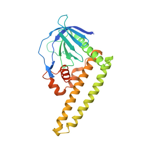
The PHCCEx domain of Tiam1/2 is a novel protein- and membrane-binding module

Terawaki, S.Kitano, K.Mori, T.Zhai, Y.Higuchi, Y.Itoh, N.Watanabe, T.Kaibuchi, K.Hakoshima, T.

(2010) EMBO J 29: 236-250

  • DOI: https://doi.org/10.1038/emboj.2009.323
  • Primary Citation of Related Structures:  
    3A8N, 3A8P, 3A8Q

  • PubMed Abstract: 

    Tiam1 and Tiam2 (Tiam1/2) are guanine nucleotide-exchange factors that possess the PH-CC-Ex (pleckstrin homology, coiled coil and extra) region that mediates binding to plasma membranes and signalling proteins in the activation of Rac GTPases. Crystal structures of the PH-CC-Ex regions revealed a single globular domain, PHCCEx domain, comprising a conventional PH subdomain associated with an antiparallel coiled coil of CC subdomain and a novel three-helical globular Ex subdomain. The PH subdomain resembles the beta-spectrin PH domain, suggesting non-canonical phosphatidylinositol binding. Mutational and binding studies indicated that CC and Ex subdomains form a positively charged surface for protein binding. We identified two unique acidic sequence motifs in Tiam1/2-interacting proteins for binding to PHCCEx domain, Motif-I in CD44 and ephrinB's and the NMDA receptor, and Motif-II in Par3 and JIP2. Our results suggest the molecular basis by which the Tiam1/2 PHCCEx domain facilitates dual binding to membranes and signalling proteins.


  • Organizational Affiliation

    Structural Biology Laboratory, Nara Institute of Science and Technology, Takayama, Ikoma, Nara, Japan.


Macromolecules
Find similar proteins by:  (by identity cutoff)  |  3D Structure
Entity ID: 1
MoleculeChains Sequence LengthOrganismDetailsImage
T-lymphoma invasion and metastasis-inducing protein 2
A, B, C, D
263Mus musculusMutation(s): 0 
Gene Names: Tiam2Kiaa2016Stef
UniProt
Find proteins for Q6ZPF3 (Mus musculus)
Explore Q6ZPF3 
Go to UniProtKB:  Q6ZPF3
Entity Groups  
Sequence Clusters30% Identity50% Identity70% Identity90% Identity95% Identity100% Identity
UniProt GroupQ6ZPF3
Sequence Annotations
Expand
  • Reference Sequence
Experimental Data & Validation

Experimental Data

  • Method: X-RAY DIFFRACTION
  • Resolution: 3.20 Å
  • R-Value Free: 0.332 
  • R-Value Work: 0.283 
  • R-Value Observed: 0.283 
  • Space Group: P 43 21 2
Unit Cell:
Length ( Å )Angle ( ˚ )
a = 105.56α = 90
b = 105.56β = 90
c = 287.58γ = 90
Software Package:
Software NamePurpose
HKL-2000data collection
SHARPphasing
CNSrefinement
HKL-2000data reduction
SCALEPACKdata scaling

Structure Validation

View Full Validation Report


Entry History 

Deposition Data

Revision History  (Full details and data files)

  • Version 1.0: 2009-11-24
    Type: Initial release
  • Version 1.1: 2011-07-13
    Changes: Version format compliance
  • Version 1.2: 2024-03-13
    Changes: Data collection, Database references