3A8G

Crystal structure of Nitrile Hydratase mutant S113A complexed with Trimethylacetonitrile


Experimental Data Snapshot

  • Method: X-RAY DIFFRACTION
  • Resolution: 1.11 Å
  • R-Value Free: 0.169 
  • R-Value Observed: 0.142 

wwPDB Validation   3D Report Full Report


This is version 1.3 of the entry. See complete history


Literature

Kinetic and structural studies on roles of the serine ligand and a strictly conserved tyrosine residue in nitrile hydratase

Yamanaka, Y.Hashimoto, K.Ohtaki, A.Noguchi, K.Yohda, M.Odaka, M.

(2010) J Biol Inorg Chem 15: 655-665

  • DOI: https://doi.org/10.1007/s00775-010-0632-3
  • Primary Citation of Related Structures:  
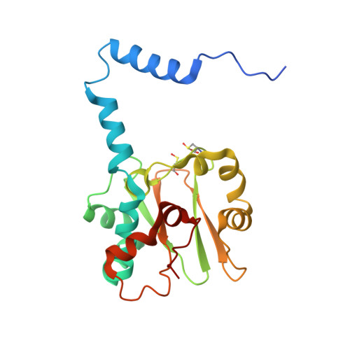
    3A8G, 3A8H, 3A8L, 3A8M, 3A8O

  • PubMed Abstract: 

    Nitrile hydratases (NHase), which catalyze the hydration of nitriles to amides, have an unusual Fe(3+) or Co(3+) center with two modified Cys ligands: cysteine sulfininate (Cys-SO(2) (-)) and either cysteine sulfenic acid or cysteine sulfenate [Cys-SO(H)]. Two catalytic mechanisms have been proposed. One is that the sulfenyl oxygen activates a water molecule, enabling nucleophilic attack on the nitrile carbon. The other is that the Ser ligand ionizes the strictly conserved Tyr, activating a water molecule. Here, we characterized mutants of Fe-type NHase from Rhodococcus erythropolis N771, replacing the Ser and Tyr residues, alphaS113A and betaY72F. The alphaS113A mutation partially affected catalytic activity and did not change the pH profiles of the kinetic parameters. UV-vis absorption spectra indicated that the electronic state of the Fe center was altered by the alphaS113A mutation, but the changes could be prevented by a competitive inhibitor, n-butyric acid. The overall structure of the alphaS113A mutant was similar to that of the wild type, but significant changes were observed around the catalytic cavity. Like the UV-vis spectra, the changes were compensated by the substrate or product. The Ser ligand is important for the structure around the catalytic cavity, but is not essential for catalysis. The betaY72F mutant exhibited no activity. The structure of the betaY72F mutant was highly conserved but was found to be the inactivated state, with alphaCys114-SO(H) oxidized to Cys-SO(2) (-), suggesting that betaTyr72 affected the electronic state of the Fe center. The catalytic mechanism is discussed on the basis of the results obtained.


  • Organizational Affiliation

    Department of Biotechnology and Life Science, Tokyo University of Agriculture and Technology, Naka-cho, Koganei, Japan.


Macromolecules
Find similar proteins by:  (by identity cutoff)  |  3D Structure
Entity ID: 1
MoleculeChains Sequence LengthOrganismDetailsImage
Nitrile hydratase subunit alpha207Rhodococcus erythropolisMutation(s): 1 
EC: 4.2.1.84
UniProt
Find proteins for P13448 (Rhodococcus erythropolis)
Explore P13448 
Go to UniProtKB:  P13448
Entity Groups  
Sequence Clusters30% Identity50% Identity70% Identity90% Identity95% Identity100% Identity
UniProt GroupP13448
Sequence Annotations
Expand
  • Reference Sequence
Find similar proteins by:  (by identity cutoff)  |  3D Structure
Entity ID: 2
MoleculeChains Sequence LengthOrganismDetailsImage
Nitrile hydratase subunit beta212Rhodococcus erythropolisMutation(s): 0 
EC: 4.2.1.84
UniProt
Find proteins for P13449 (Rhodococcus erythropolis)
Explore P13449 
Go to UniProtKB:  P13449
Entity Groups  
Sequence Clusters30% Identity50% Identity70% Identity90% Identity95% Identity100% Identity
UniProt GroupP13449
Sequence Annotations
Expand
  • Reference Sequence
Small Molecules
Modified Residues  2 Unique
IDChains TypeFormula2D DiagramParent
CSD
Query on CSD
A
L-PEPTIDE LINKINGC3 H7 N O4 SCYS
CSO
Query on CSO
A
L-PEPTIDE LINKINGC3 H7 N O3 SCYS
Experimental Data & Validation

Experimental Data

  • Method: X-RAY DIFFRACTION
  • Resolution: 1.11 Å
  • R-Value Free: 0.169 
  • R-Value Observed: 0.142 
  • Space Group: C 1 2 1
Unit Cell:
Length ( Å )Angle ( ˚ )
a = 114.411α = 90
b = 60.268β = 124.92
c = 81.81γ = 90
Software Package:
Software NamePurpose
HKL-2000data collection
MOLREPphasing
REFMACrefinement
HKL-2000data reduction
HKL-2000data scaling
SHELXphasing
SHELXL-97refinement

Structure Validation

View Full Validation Report


Entry History 

Deposition Data

Revision History  (Full details and data files)

  • Version 1.0: 2010-04-14
    Type: Initial release
  • Version 1.1: 2011-07-13
    Changes: Version format compliance
  • Version 1.2: 2014-01-22
    Changes: Database references
  • Version 1.3: 2021-11-10
    Changes: Database references, Derived calculations