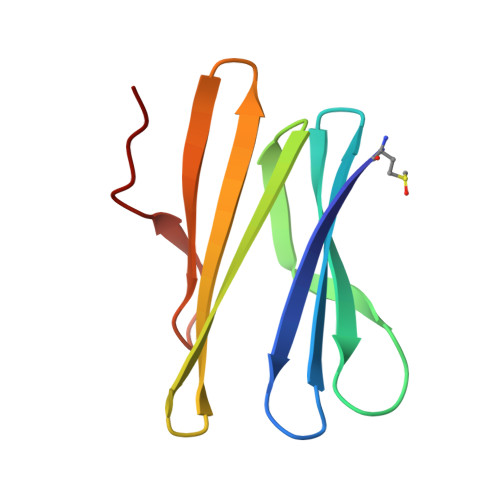
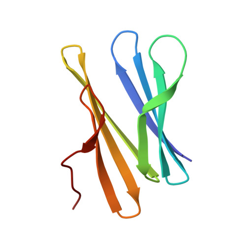
3ZW1

Structure of Bambl lectine in complex with lewix x antigen


Experimental Data Snapshot

  • Method: X-RAY DIFFRACTION
  • Resolution: 1.60 Å
  • R-Value Free: 0.189 
  • R-Value Work: 0.153 
  • R-Value Observed: 0.155 

wwPDB Validation   3D Report Full Report


Ligand Structure Quality Assessment 


This is version 2.1 of the entry. See complete history


Literature

Deciphering the Glycan Preference of Bacterial Lectins by Glycan Array and Molecular Docking with Validation by Microcalorimetry and Crystallography.

Topin, J.Arnaud, J.Sarkar, A.Audfray, A.Gillon, E.Perez, S.Jamet, H.Varrot, A.Imberty, A.Thomas, A.

(2013) PLoS One 8: 71149

  • DOI: https://doi.org/10.1371/journal.pone.0071149
  • Primary Citation of Related Structures:  
    3ZW1

  • PubMed Abstract: 

    Recent advances in glycobiology revealed the essential role of lectins for deciphering the glycocode by specific recognition of carbohydrates. Integrated multiscale approaches are needed for characterizing lectin specificity: combining on one hand high-throughput analysis by glycan array experiments and systematic molecular docking of oligosaccharide libraries and on the other hand detailed analysis of the lectin/oligosaccharide interaction by x-ray crystallography, microcalorimetry and free energy calculations. The lectins LecB from Pseudomonas aeruginosa and BambL from Burkholderia ambifaria are part of the virulence factors used by the pathogenic bacteria to invade the targeted host. These two lectins are not related but both recognize fucosylated oligosaccharides such as the histo-blood group oligosaccharides of the ABH(O) and Lewis epitopes. The specificities were characterized using semi-quantitative data from glycan array and analyzed by molecular docking with the Glide software. Reliable prediction of protein/oligosaccharide structures could be obtained as validated by existing crystal structures of complexes. Additionally, the crystal structure of BambL/Lewis x was determined at 1.6 Å resolution, which confirms that Lewis x has to adopt a high-energy conformation so as to bind to this lectin. Free energies of binding were calculated using a procedure combining the Glide docking protocol followed by free energy rescoring with the Prime/Molecular Mechanics Generalized Born Surface Area (MM-GBSA) method. The calculated data were in reasonable agreement with experimental free energies of binding obtained by titration microcalorimetry. The established predictive protocol is proposed to rationalize large sets of data such as glycan arrays and to help in lead discovery projects based on such high throughput technology.


  • Organizational Affiliation

    CERMAV- Centre national de la recherche scientifique UPR5301 (affiliated to Université Joseph Fourier and ICMG), BP53, 38041 Grenoble, France ; Département de Chimie Moléculaire, UMR- Centre national de la recherche scientifique 5250 & ICMG FR 2607, Université Joseph Fourier, BP 53, 38041 Grenoble, France.


Macromolecules
Find similar proteins by:  (by identity cutoff)  |  3D Structure
Entity ID: 1
MoleculeChains Sequence LengthOrganismDetailsImage
BAMBL LECTIN87Burkholderia ambifariaMutation(s): 0 
UniProt
Find proteins for Q0B4G1 (Burkholderia ambifaria (strain ATCC BAA-244 / AMMD))
Explore Q0B4G1 
Go to UniProtKB:  Q0B4G1
Entity Groups  
Sequence Clusters30% Identity50% Identity70% Identity90% Identity95% Identity100% Identity
UniProt GroupQ0B4G1
Sequence Annotations
Expand
  • Reference Sequence
Find similar proteins by:  (by identity cutoff)  |  3D Structure
Entity ID: 2
MoleculeChains Sequence LengthOrganismDetailsImage
BAMBL LECTIN87Burkholderia ambifariaMutation(s): 0 
UniProt
Find proteins for Q0B4G1 (Burkholderia ambifaria (strain ATCC BAA-244 / AMMD))
Explore Q0B4G1 
Go to UniProtKB:  Q0B4G1
Entity Groups  
Sequence Clusters30% Identity50% Identity70% Identity90% Identity95% Identity100% Identity
UniProt GroupQ0B4G1
Sequence Annotations
Expand
  • Reference Sequence
Oligosaccharides

Help

Entity ID: 3
MoleculeChains Length2D Diagram Glycosylation3D Interactions
alpha-L-fucopyranose-(1-3)-2-acetamido-2-deoxy-beta-D-glucopyranose
C, F
2N/A
Glycosylation Resources
GlyTouCan:  G80587NA
GlyCosmos:  G80587NA
GlyGen:  G80587NA
Entity ID: 4
MoleculeChains Length2D Diagram Glycosylation3D Interactions
alpha-L-fucopyranose-(1-3)-[beta-D-galactopyranose-(1-4)]2-acetamido-2-deoxy-beta-D-glucopyranose-(1-3)-beta-D-galactopyranose
D, E
4N/A
Glycosylation Resources
GlyTouCan:  G81345OW
GlyCosmos:  G81345OW
GlyGen:  G81345OW
Small Molecules
Ligands 1 Unique
IDChains Name / Formula / InChI Key2D Diagram3D Interactions
GLA
Query on GLA

Download Ideal Coordinates CCD File 
G [auth B]alpha-D-galactopyranose
C6 H12 O6
WQZGKKKJIJFFOK-PHYPRBDBSA-N
Modified Residues  1 Unique
IDChains TypeFormula2D DiagramParent
MHO
Query on MHO
A
L-PEPTIDE LINKINGC5 H11 N O3 SMET
Experimental Data & Validation

Experimental Data

  • Method: X-RAY DIFFRACTION
  • Resolution: 1.60 Å
  • R-Value Free: 0.189 
  • R-Value Work: 0.153 
  • R-Value Observed: 0.155 
  • Space Group: P 21 3
Unit Cell:
Length ( Å )Angle ( ˚ )
a = 81.44α = 90
b = 81.44β = 90
c = 81.44γ = 90
Software Package:
Software NamePurpose
REFMACrefinement
MOSFLMdata reduction
SCALAdata scaling
PHASERphasing

Structure Validation

View Full Validation Report


Ligand Structure Quality Assessment 


Entry History 

Deposition Data

Revision History  (Full details and data files)

  • Version 1.0: 2011-11-16
    Type: Initial release
  • Version 1.1: 2013-10-09
    Changes: Database references
  • Version 2.0: 2020-07-29
    Type: Remediation
    Reason: Carbohydrate remediation
    Changes: Atomic model, Data collection, Derived calculations, Other, Structure summary
  • Version 2.1: 2023-12-20
    Changes: Data collection, Database references, Refinement description, Structure summary