Structural Characterisation of Tpx from Yersinia Pseudotuberculosis Reveals Insights Into the Binding of Salicylidene Acylhydrazide Compounds.
Gabrielsen, M., Beckham, K.S.H., Feher, V.A., Zetterstrom, C.E., Wang, D., Muller, S., Elofsson, M., Amaro, R.E., Byron, O., Roe, A.J.(2012) PLoS One 7: 32217
- PubMed: 22384182
- DOI: https://doi.org/10.1371/journal.pone.0032217
- PubMed Abstract:
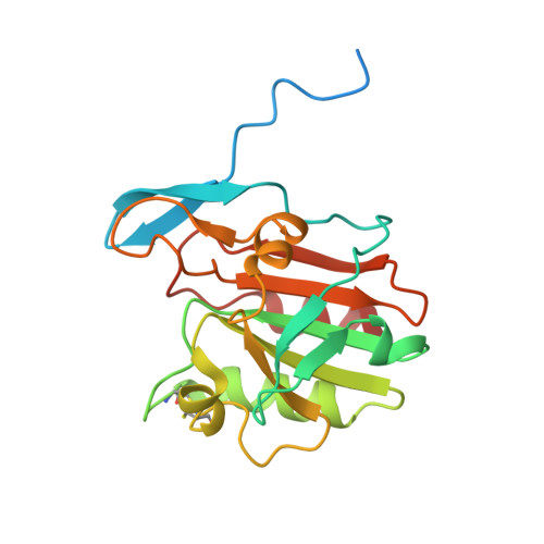
Thiol peroxidase, Tpx, has been shown to be a target protein of the salicylidene acylhydrazide class of antivirulence compounds. In this study we present the crystal structures of Tpx from Y. pseudotuberculosis (ypTpx) in the oxidised and reduced states, together with the structure of the C61S mutant. The structures solved are consistent with previously solved atypical 2-Cys thiol peroxidases, including that for "forced" reduced states using the C61S mutant. In addition, by investigating the solution structure of ypTpx using small angle X-ray scattering (SAXS), we have confirmed that reduced state ypTpx in solution is a homodimer. The solution structure also reveals flexibility around the dimer interface. Notably, the conformational changes observed between the redox states at the catalytic triad and at the dimer interface have implications for substrate and inhibitor binding. The structural data were used to model the binding of two salicylidene acylhydrazide compounds to the oxidised structure of ypTpx. Overall, the study provides insights into the binding of the salicylidene acylhydrazides to ypTpx, aiding our long-term strategy to understand the mode of action of this class of compounds.
Organizational Affiliation:
Institute of Infection, Immunity and Immunology, College of Medical, Veterinary and Life Sciences, University of Glasgow, Glasgow, United Kingdom.