3ZIZ

Crystal structure of Podospora anserina GH5 beta-(1,4)-mannanase


Experimental Data Snapshot

  • Method: X-RAY DIFFRACTION
  • Resolution: 1.40 Å
  • R-Value Free: 0.172 
  • R-Value Work: 0.150 
  • R-Value Observed: 0.151 

wwPDB Validation   3D Report Full Report


This is version 1.2 of the entry. See complete history


Literature

Structural and Biochemical Analyses of Glycoside Hydrolase Families 5 and 26 Beta-(1,4)-Mannanases from Podospora Anserina Reveal Differences Upon Manno-Oligosaccharides Catalysis.

Couturier, M.Roussel, A.Rosengren, A.Leone, P.Stalbrand, H.Berrin, J.G.

(2013) J Biol Chem 288: 14624

  • DOI: https://doi.org/10.1074/jbc.M113.459438
  • Primary Citation of Related Structures:  
    3ZIZ, 3ZM8

  • PubMed Abstract: 

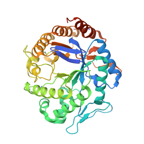
    The microbial deconstruction of the plant cell wall is a key biological process that is of increasing importance with the development of a sustainable biofuel industry. The glycoside hydrolase families GH5 (PaMan5A) and GH26 (PaMan26A) endo-β-1,4-mannanases from the coprophilic ascomycete Podospora anserina contribute to the enzymatic degradation of lignocellulosic biomass. In this study, P. anserina mannanases were further subjected to detailed comparative analysis of their substrate specificities, active site organization, and transglycosylation capacity. Although PaMan5A displays a classical mode of action, PaMan26A revealed an atypical hydrolysis pattern with the release of mannotetraose and mannose from mannopentaose resulting from a predominant binding mode involving the -4 subsite. The crystal structures of PaMan5A and PaMan26A were solved at 1.4 and 2.85 Å resolution, respectively. Analysis of the PaMan26A structure supported strong interaction with substrate at the -4 subsite mediated by two aromatic residues Trp-244 and Trp-245. The PaMan26A structure appended to its family 35 carbohydrate binding module revealed a short and proline-rich rigid linker that anchored together the catalytic and the binding modules.


  • Organizational Affiliation

    INRA, UMR1163 BCF, Aix Marseille Université, Polytech Marseille, F-13288 Marseille, France.


Macromolecules
Find similar proteins by:  (by identity cutoff)  |  3D Structure
Entity ID: 1
MoleculeChains Sequence LengthOrganismDetailsImage
GH5 ENDO-BETA-1,4-MANNANASE382Podospora anserinaMutation(s): 0 
EC: 3.2.1.78
UniProt
Find proteins for B2B3C0 (Podospora anserina (strain S / ATCC MYA-4624 / DSM 980 / FGSC 10383))
Explore B2B3C0 
Go to UniProtKB:  B2B3C0
Entity Groups  
Sequence Clusters30% Identity50% Identity70% Identity90% Identity95% Identity100% Identity
UniProt GroupB2B3C0
Sequence Annotations
Expand
  • Reference Sequence
Small Molecules
Ligands 2 Unique
IDChains Name / Formula / InChI Key2D Diagram3D Interactions
TRS
Query on TRS

Download Ideal Coordinates CCD File 
C [auth A]2-AMINO-2-HYDROXYMETHYL-PROPANE-1,3-DIOL
C4 H12 N O3
LENZDBCJOHFCAS-UHFFFAOYSA-O
GOL
Query on GOL

Download Ideal Coordinates CCD File 
B [auth A]GLYCEROL
C3 H8 O3
PEDCQBHIVMGVHV-UHFFFAOYSA-N
Experimental Data & Validation

Experimental Data

  • Method: X-RAY DIFFRACTION
  • Resolution: 1.40 Å
  • R-Value Free: 0.172 
  • R-Value Work: 0.150 
  • R-Value Observed: 0.151 
  • Space Group: P 21 21 21
Unit Cell:
Length ( Å )Angle ( ˚ )
a = 56.87α = 90
b = 57.9β = 90
c = 97.86γ = 90
Software Package:
Software NamePurpose
BUSTERrefinement
XDSdata reduction
SCALAdata scaling
AMoREphasing

Structure Validation

View Full Validation Report


Entry History 

Deposition Data

Revision History  (Full details and data files)

  • Version 1.0: 2013-04-17
    Type: Initial release
  • Version 1.1: 2013-06-12
    Changes: Database references
  • Version 1.2: 2023-12-20
    Changes: Data collection, Database references, Derived calculations, Other, Refinement description