3ZI6

Structure of thermolysin solved by SAD from data collected by Direct Data Collection (DDC) using the GRob robot goniometer


Experimental Data Snapshot

  • Method: X-RAY DIFFRACTION
  • Resolution: 2.00 Å
  • R-Value Free: 0.204 
  • R-Value Work: 0.164 
  • R-Value Observed: 0.166 

wwPDB Validation   3D Report Full Report


This is version 1.3 of the entry. See complete history


Literature

Recent Progress in Robot-Based Systems for Crystallography and Their Contribution to Drug Discovery.

Ferrer, J.L.Larive, N.A.Bowler, M.W.Nurizzo, D.

(2013) Expert Opin Drug Discov 8: 835

  • DOI: https://doi.org/10.1517/17460441.2013.793666
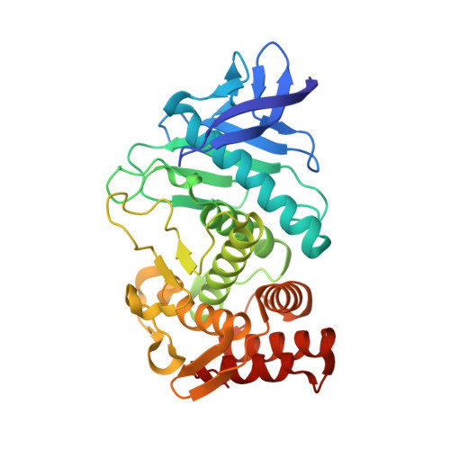
  • Primary Citation of Related Structures:  
    3ZI6, 3ZI7

  • PubMed Abstract: 

    X-ray crystallography is the main tool for macromolecular structure solution at atomic resolution. It provides key information for the understanding of protein function, opening opportunities for the modulation of enzymatic mechanisms, and protein-ligand interactions. As a consequence, macromolecular crystallography plays an essential role in drug design, as well as in the a posteriori validation of drug mechanisms.


  • Organizational Affiliation

    Commissariat à l'Energie Atomique et aux Energies Alternatives (CEA), Centre National de la Recherche Scientifique (CNRS), Université Joseph Fourier (UJF), Institut de Biologie Structurale Jean-Pierre Ebel (IBS), F-38027 Grenoble Cedex 1, France. jean-luc.ferrer@ibs.fr


Macromolecules
Find similar proteins by:  (by identity cutoff)  |  3D Structure
Entity ID: 1
MoleculeChains Sequence LengthOrganismDetailsImage
THERMOLYSIN316Bacillus thermoproteolyticusMutation(s): 0 
EC: 3.4.24.27
UniProt
Find proteins for P00800 (Bacillus thermoproteolyticus)
Explore P00800 
Go to UniProtKB:  P00800
Entity Groups  
Sequence Clusters30% Identity50% Identity70% Identity90% Identity95% Identity100% Identity
UniProt GroupP00800
Sequence Annotations
Expand
  • Reference Sequence
Small Molecules
Ligands 4 Unique
IDChains Name / Formula / InChI Key2D Diagram3D Interactions
LYS
Query on LYS

Download Ideal Coordinates CCD File 
C [auth A]LYSINE
C6 H15 N2 O2
KDXKERNSBIXSRK-YFKPBYRVSA-O
VAL
Query on VAL

Download Ideal Coordinates CCD File 
B [auth A]VALINE
C5 H11 N O2
KZSNJWFQEVHDMF-BYPYZUCNSA-N
ZN
Query on ZN

Download Ideal Coordinates CCD File 
D [auth A]ZINC ION
Zn
PTFCDOFLOPIGGS-UHFFFAOYSA-N
CA
Query on CA

Download Ideal Coordinates CCD File 
E [auth A],
F [auth A],
G [auth A],
H [auth A]
CALCIUM ION
Ca
BHPQYMZQTOCNFJ-UHFFFAOYSA-N
Experimental Data & Validation

Experimental Data

  • Method: X-RAY DIFFRACTION
  • Resolution: 2.00 Å
  • R-Value Free: 0.204 
  • R-Value Work: 0.164 
  • R-Value Observed: 0.166 
  • Space Group: P 61 2 2
Unit Cell:
Length ( Å )Angle ( ˚ )
a = 92.754α = 90
b = 92.754β = 90
c = 129.224γ = 120
Software Package:
Software NamePurpose
REFMACrefinement
MOSFLMdata reduction
SCALAdata scaling
SHELXCDEphasing

Structure Validation

View Full Validation Report


Entry History 

Deposition Data

  • Released Date: 2013-05-01 
  • Deposition Author(s): Bowler, M.W.

Revision History  (Full details and data files)

  • Version 1.0: 2013-05-01
    Type: Initial release
  • Version 1.1: 2013-05-22
    Changes: Database references, Refinement description, Structure summary
  • Version 1.2: 2013-07-03
    Changes: Database references
  • Version 1.3: 2018-06-20
    Changes: Advisory, Data collection, Derived calculations