3ZH4

crystal structure of S. pneumoniae Hungary 19A MurA1 in complex with citrate


Experimental Data Snapshot

  • Method: X-RAY DIFFRACTION
  • Resolution: 1.80 Å
  • R-Value Free: 0.204 
  • R-Value Work: 0.160 
  • R-Value Observed: 0.163 

wwPDB Validation   3D Report Full Report


This is version 1.2 of the entry. See complete history


Literature

Heteroresistance to Fosfomycin is Predominant in Streptococcus Pneumoniae and Depends on Mura1 Gene.

Engel, H.Gutierrez-Fernandez, J.Fluckiger, C.Martinez-Ripoll, M.Muhlemann, K.Hermoso, J.A.Hilty, M.Hathaway, L.J.

(2013) Antimicrob Agents Chemother 57: 2801

  • DOI: https://doi.org/10.1128/AAC.00223-13
  • Primary Citation of Related Structures:  
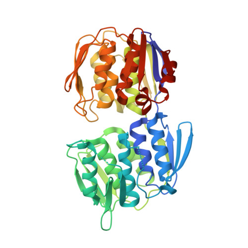
    3ZH3, 3ZH4

  • PubMed Abstract: 

    Fosfomycin targets the first step of peptidoglycan biosynthesis in Streptococcus pneumoniae catalyzed by UDP-N-acetylglucosamine enolpyruvyltransferase (MurA1). We investigated whether heteroresistance to fosfomycin occurs in S. pneumoniae. We found that of 11 strains tested, all but 1 (Hungary(19A)) displayed heteroresistance and that deletion of murA1 abolished heteroresistance. Hungary(19A) differs from the other strains by a single amino acid substitution in MurA1 (Ala(364)Thr). To test whether this substitution is responsible for the lack of heteroresistance, it was introduced into strain D39. The heteroresistance phenotype of strain D39 was not changed. Furthermore, no relevant structural differences between the MurA1 crystal structures of heteroresistant strain D39 and nonheteroresistant strain Hungary(19A) were found. Our results reveal that heteroresistance to fosfomycin is the predominant phenotype of S. pneumoniae and that MurA1 is required for heteroresistance to fosfomycin but is not the only factor involved. The findings provide a caveat for any future use of fosfomycin in the treatment of pneumococcal infections.


  • Organizational Affiliation

    Institute for Infectious Diseases, Faculty of Medicine, University of Bern, Bern, Switzerland.


Macromolecules
Find similar proteins by:  (by identity cutoff)  |  3D Structure
Entity ID: 1
MoleculeChains Sequence LengthOrganismDetailsImage
UDP-N-ACETYLGLUCOSAMINE 1-CARBOXYVINYLTRANSFERASE419Streptococcus pneumoniaeMutation(s): 0 
EC: 2.5.1.7
UniProt
Find proteins for B1IBM3 (Streptococcus pneumoniae (strain Hungary19A-6))
Explore B1IBM3 
Go to UniProtKB:  B1IBM3
Entity Groups  
Sequence Clusters30% Identity50% Identity70% Identity90% Identity95% Identity100% Identity
UniProt GroupB1IBM3
Sequence Annotations
Expand
  • Reference Sequence
Small Molecules
Ligands 2 Unique
IDChains Name / Formula / InChI Key2D Diagram3D Interactions
FLC
Query on FLC

Download Ideal Coordinates CCD File 
B [auth A]CITRATE ANION
C6 H5 O7
KRKNYBCHXYNGOX-UHFFFAOYSA-K
PEG
Query on PEG

Download Ideal Coordinates CCD File 
C [auth A]DI(HYDROXYETHYL)ETHER
C4 H10 O3
MTHSVFCYNBDYFN-UHFFFAOYSA-N
Experimental Data & Validation

Experimental Data

  • Method: X-RAY DIFFRACTION
  • Resolution: 1.80 Å
  • R-Value Free: 0.204 
  • R-Value Work: 0.160 
  • R-Value Observed: 0.163 
  • Space Group: P 21 21 2
Unit Cell:
Length ( Å )Angle ( ˚ )
a = 106.584α = 90
b = 70.36β = 90
c = 45.241γ = 90
Software Package:
Software NamePurpose
PHENIXrefinement
XDSdata reduction
SCALAdata scaling
PHASERphasing

Structure Validation

View Full Validation Report


Entry History 

Deposition Data

Revision History  (Full details and data files)

  • Version 1.0: 2013-04-24
    Type: Initial release
  • Version 1.1: 2013-05-29
    Changes: Database references
  • Version 1.2: 2023-12-20
    Changes: Data collection, Database references, Derived calculations, Other, Refinement description