3ZG5

Crystal structure of PBP2a from MRSA in complex with peptidoglycan analogue at allosteric


Experimental Data Snapshot

  • Method: X-RAY DIFFRACTION
  • Resolution: 2.55 Å
  • R-Value Free: 0.290 
  • R-Value Work: 0.214 
  • R-Value Observed: 0.217 

wwPDB Validation   3D Report Full Report


Ligand Structure Quality Assessment 


This is version 2.2 of the entry. See complete history


Literature

How Allosteric Control of Staphylococcus Aureus Penicillin Binding Protein 2A Enables Methicillin Resistance and Physiological Function

Otero, L.H.Rojas-Altuve, A.Llarrull, L.I.Carrasco-Lopez, C.Kumarasiri, M.Lastochkin, E.Fishovitz, J.Dawley, M.Hesek, D.Lee, M.Johnson, J.W.Fisher, J.F.Chang, M.Mobashery, S.Hermoso, J.A.

(2013) Proc Natl Acad Sci U S A 110: 16808

  • DOI: https://doi.org/10.1073/pnas.1300118110
  • Primary Citation of Related Structures:  
    3ZG5

  • PubMed Abstract: 

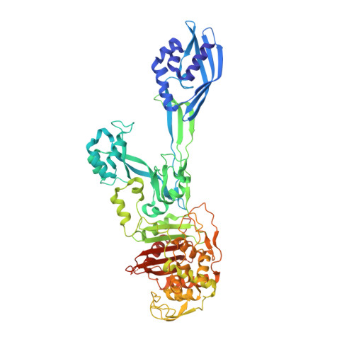
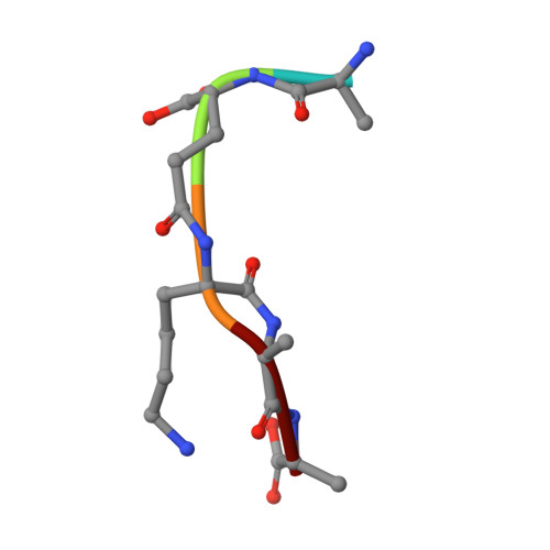
    The expression of penicillin binding protein 2a (PBP2a) is the basis for the broad clinical resistance to the β-lactam antibiotics by methicillin-resistant Staphylococcus aureus (MRSA). The high-molecular mass penicillin binding proteins of bacteria catalyze in separate domains the transglycosylase and transpeptidase activities required for the biosynthesis of the peptidoglycan polymer that comprises the bacterial cell wall. In bacteria susceptible to β-lactam antibiotics, the transpeptidase activity of their penicillin binding proteins (PBPs) is lost as a result of irreversible acylation of an active site serine by the β-lactam antibiotics. In contrast, the PBP2a of MRSA is resistant to β-lactam acylation and successfully catalyzes the DD-transpeptidation reaction necessary to complete the cell wall. The inability to contain MRSA infection with β-lactam antibiotics is a continuing public health concern. We report herein the identification of an allosteric binding domain--a remarkable 60 Å distant from the DD-transpeptidase active site--discovered by crystallographic analysis of a soluble construct of PBP2a. When this allosteric site is occupied, a multiresidue conformational change culminates in the opening of the active site to permit substrate entry. This same crystallographic analysis also reveals the identity of three allosteric ligands: muramic acid (a saccharide component of the peptidoglycan), the cell wall peptidoglycan, and ceftaroline, a recently approved anti-MRSA β-lactam antibiotic. The ability of an anti-MRSA β-lactam antibiotic to stimulate allosteric opening of the active site, thus predisposing PBP2a to inactivation by a second β-lactam molecule, opens an unprecedented realm for β-lactam antibiotic structure-based design.


  • Organizational Affiliation

    Departamento de Cristalografía y Biología Estructural, Instituto de Química-Física "Rocasolano," Consejo Superior de Investigaciones Cientificas, 28006 Madrid, Spain.


Macromolecules
Find similar proteins by:  (by identity cutoff)  |  3D Structure
Entity ID: 1
MoleculeChains Sequence LengthOrganismDetailsImage
Penicillin binding protein 2 prime
A, B
642Staphylococcus aureus subsp. aureus Mu50Mutation(s): 0 
Gene Names: mecASAV0041
UniProt
Find proteins for A0A0H3JPA5 (Staphylococcus aureus (strain Mu50 / ATCC 700699))
Explore A0A0H3JPA5 
Go to UniProtKB:  A0A0H3JPA5
Entity Groups  
Sequence Clusters30% Identity50% Identity70% Identity90% Identity95% Identity100% Identity
UniProt GroupA0A0H3JPA5
Sequence Annotations
Expand
  • Reference Sequence

Find similar proteins by:  Sequence   |   3D Structure  

Entity ID: 2
MoleculeChains Sequence LengthOrganismDetailsImage
PEPTIDOGLYCAN ANALOGUE
C, D
5synthetic constructMutation(s): 0 
Sequence Annotations
Expand
  • Reference Sequence
Small Molecules
Ligands 3 Unique
IDChains Name / Formula / InChI Key2D Diagram3D Interactions
AMV
Query on AMV

Download Ideal Coordinates CCD File 
M [auth C],
N [auth D]
methyl 2-acetamido-3-O-[(1R)-1-carboxyethyl]-2-deoxy-beta-D-glucopyranoside
C12 H21 N O8
UXEQYDNCHCKBIL-PKKPQKKZSA-N
CD
Query on CD

Download Ideal Coordinates CCD File 
E [auth A],
G [auth A],
I [auth B],
J [auth B]
CADMIUM ION
Cd
WLZRMCYVCSSEQC-UHFFFAOYSA-N
CL
Query on CL

Download Ideal Coordinates CCD File 
F [auth A],
H [auth A],
K [auth B],
L [auth B]
CHLORIDE ION
Cl
VEXZGXHMUGYJMC-UHFFFAOYSA-M
Experimental Data & Validation

Experimental Data

  • Method: X-RAY DIFFRACTION
  • Resolution: 2.55 Å
  • R-Value Free: 0.290 
  • R-Value Work: 0.214 
  • R-Value Observed: 0.217 
  • Space Group: P 21 21 21
Unit Cell:
Length ( Å )Angle ( ˚ )
a = 80.54α = 90
b = 102.02β = 90
c = 185.98γ = 90
Software Package:
Software NamePurpose
PHENIXrefinement
PHASERphasing

Structure Validation

View Full Validation Report


Ligand Structure Quality Assessment 


Entry History 

Deposition Data

Revision History  (Full details and data files)

  • Version 1.0: 2013-10-09
    Type: Initial release
  • Version 1.1: 2013-10-30
    Changes: Database references
  • Version 1.2: 2016-12-14
    Changes: Source and taxonomy
  • Version 1.3: 2020-07-29
    Type: Remediation
    Reason: Carbohydrate remediation
    Changes: Advisory, Derived calculations, Other, Structure summary
  • Version 2.0: 2022-12-07
    Changes: Advisory, Atomic model, Data collection, Database references, Derived calculations, Polymer sequence, Source and taxonomy, Structure summary
  • Version 2.1: 2023-07-05
    Changes: Advisory, Derived calculations
  • Version 2.2: 2024-02-07
    Changes: Data collection, Refinement description