The Hydrogen Peroxide Induced Radical Behaviour in Human Cytochrome C Phospholipid Complexes: Implications for the Enhanced Pro-Apoptotic Activity of the G41S Mutant
Rajagopal, B.S., Edzuma, A.N., Hough, M.A., Blundell, K.L.I.M., Kagan, V.E., Kapralov, A.A., Fraser, L.A., Butt, J.N., Silkstone, G.G., Wilson, M.T., Svistunenko, D.A., Worrall, J.A.R.(2013) Biochem J 456: 441
- PubMed: 24099549
- DOI: https://doi.org/10.1042/BJ20130758
- Primary Citation of Related Structures:
3ZCF, 3ZOO - PubMed Abstract:
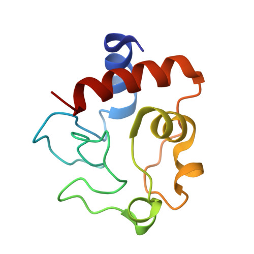
We have investigated whether the pro-apoptotic properties of the G41S mutant of human cytochrome c can be explained by a higher than wild-type peroxidase activity triggered by phospholipid binding. A key complex in mitochondrial apoptosis involves cytochrome c and the phospholipid cardiolipin. In this complex cytochrome c has its native axial Met(80) ligand dissociated from the haem-iron, considerably augmenting the peroxidase capability of the haem group upon H2O2 binding. By EPR spectroscopy we reveal that the magnitude of changes in the paramagnetic haem states, as well as the yield of protein-bound free radical, is dependent on the phospholipid used and is considerably greater in the G41S mutant. A high-resolution X-ray crystal structure of human cytochrome c was determined and, in combination with the radical EPR signal analysis, two tyrosine residues, Tyr(46) and Tyr(48), have been rationalized to be putative radical sites. Subsequent single and double tyrosine-to-phenylalanine mutations revealed that the EPR signal of the radical, found to be similar in all variants, including G41S and wild-type, originates not from a single tyrosine residue, but is instead a superimposition of multiple EPR signals from different radical sites. We propose a mechanism of multiple radical formations in the cytochrome c-phospholipid complexes under H2O2 treatment, consistent with the stabilization of the radical in the G41S mutant, which elicits a greater peroxidase activity from cytochrome c and thus has implications in mitochondrial apoptosis.
Organizational Affiliation:
*School of Biological Sciences, University of Essex, Wivenhoe Park, Colchester, Essex CO4 3SQ, U.K.