3WKX

Crystal structure of GH127 beta-L-arabinofuranosidase HypBA1 from Bifidobacterium longum arabinose complex form


Experimental Data Snapshot

  • Method: X-RAY DIFFRACTION
  • Resolution: 2.00 Å
  • R-Value Free: 0.279 
  • R-Value Work: 0.226 
  • R-Value Observed: 0.229 

wwPDB Validation   3D Report Full Report


Ligand Structure Quality Assessment 


This is version 2.1 of the entry. See complete history


Literature

Crystal structure of glycoside hydrolase family 127 beta-l-arabinofuranosidase from Bifidobacterium longum.

Ito, T.Saikawa, K.Kim, S.Fujita, K.Ishiwata, A.Kaeothip, S.Arakawa, T.Wakagi, T.Beckham, G.T.Ito, Y.Fushinobu, S.

(2014) Biochem Biophys Res Commun 447: 32-37

  • DOI: https://doi.org/10.1016/j.bbrc.2014.03.096
  • Primary Citation of Related Structures:  
    3WKW, 3WKX

  • PubMed Abstract: 

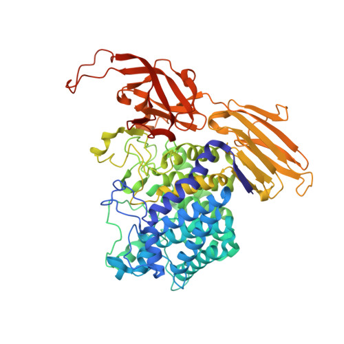
    Enzymes acting on β-linked arabinofuranosides have been unknown until recently, in spite of wide distribution of β-l-arabinofuranosyl oligosaccharides in plant cells. Recently, a β-l-arabinofuranosidase from the glycoside hydrolase family 127 (HypBA1) was discovered in the newly characterized degradation system of hydroxyproline-linked β-l-arabinooligosaccharides in the bacterium Bifidobacterium longum. Here, we report the crystal structure of HypBA1 in the ligand-free and β-l-arabinofuranose complex forms. The structure of HypBA1 consists of a catalytic barrel domain and two additional β-sandwich domains, with one β-sandwich domain involved in the formation of a dimer. Interestingly, there is an unprecedented metal-binding motif with Zn(2+) coordinated by glutamate and three cysteines in the active site. The glutamate residue is located far from the anomeric carbon of the β-l-arabinofuranose ligand, but one cysteine residue is appropriately located for nucleophilic attack for glycosidic bond cleavage. The residues around the active site are highly conserved among GH127 members. Based on biochemical experiments and quantum mechanical calculations, a possible reaction mechanism involving cysteine as the nucleophile is proposed.


  • Organizational Affiliation

    Department of Biotechnology, The University of Tokyo, Tokyo, Japan.


Macromolecules
Find similar proteins by:  (by identity cutoff)  |  3D Structure
Entity ID: 1
MoleculeChains Sequence LengthOrganismDetailsImage
Non-reducing end beta-L-arabinofuranosidase669Bifidobacterium longum subsp. longum JCM 1217Mutation(s): 0 
Gene Names: hypBA1
EC: 3.2.1.185
UniProt
Find proteins for E8MGH8 (Bifidobacterium longum subsp. longum (strain ATCC 15707 / DSM 20219 / JCM 1217 / NCTC 11818 / E194b))
Explore E8MGH8 
Go to UniProtKB:  E8MGH8
Entity Groups  
Sequence Clusters30% Identity50% Identity70% Identity90% Identity95% Identity100% Identity
UniProt GroupE8MGH8
Sequence Annotations
Expand
  • Reference Sequence
Small Molecules
Experimental Data & Validation

Experimental Data

  • Method: X-RAY DIFFRACTION
  • Resolution: 2.00 Å
  • R-Value Free: 0.279 
  • R-Value Work: 0.226 
  • R-Value Observed: 0.229 
  • Space Group: P 32 2 1
Unit Cell:
Length ( Å )Angle ( ˚ )
a = 77.726α = 90
b = 77.726β = 90
c = 252.668γ = 120
Software Package:
Software NamePurpose
HKL-2000data collection
PHENIXmodel building
REFMACrefinement
HKL-2000data reduction
HKL-2000data scaling
PHENIXphasing

Structure Validation

View Full Validation Report


Ligand Structure Quality Assessment 


Entry History 

Deposition Data

Revision History  (Full details and data files)

  • Version 1.0: 2014-04-30
    Type: Initial release
  • Version 2.0: 2020-07-29
    Type: Remediation
    Reason: Carbohydrate remediation
    Changes: Atomic model, Data collection, Database references, Derived calculations
  • Version 2.1: 2022-08-24
    Changes: Database references, Structure summary