3WDN

High-resolution X-ray crystal structure of bovine H-protein using a high-pressure cryocooling method


Experimental Data Snapshot

  • Method: X-RAY DIFFRACTION
  • Resolution: 0.86 Å
  • R-Value Free: 0.139 
  • R-Value Work: 0.114 

wwPDB Validation   3D Report Full Report


This is version 1.2 of the entry. See complete history


Literature

High-resolution X-ray crystal structure of bovine H-protein using the high-pressure cryocooling method

Higashiura, A.Ohta, K.Masaki, M.Sato, M.Inaka, K.Tanaka, H.Nakagawa, A.

(2013) J Synchrotron Radiat 20: 989-993

  • DOI: https://doi.org/10.1107/S090904951302373X
  • Primary Citation of Related Structures:  
    3WDN

  • PubMed Abstract: 

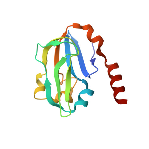
    Recently, many technical improvements in macromolecular X-ray crystallography have increased the number of structures deposited in the Protein Data Bank and improved the resolution limit of protein structures. Almost all high-resolution structures have been determined using a synchrotron radiation source in conjunction with cryocooling techniques, which are required in order to minimize radiation damage. However, optimization of cryoprotectant conditions is a time-consuming and difficult step. To overcome this problem, the high-pressure cryocooling method was developed (Kim et al., 2005) and successfully applied to many protein-structure analyses. In this report, using the high-pressure cryocooling method, the X-ray crystal structure of bovine H-protein was determined at 0.86 Å resolution. Structural comparisons between high- and ambient-pressure cryocooled crystals at ultra-high resolution illustrate the versatility of this technique. This is the first ultra-high-resolution X-ray structure obtained using the high-pressure cryocooling method.


  • Organizational Affiliation

    Institute for Protein Research, Osaka University, 3-2 Yamadaoka, Suita, Osaka 565-0871, Japan.


Macromolecules
Find similar proteins by:  (by identity cutoff)  |  3D Structure
Entity ID: 1
MoleculeChains Sequence LengthOrganismDetailsImage
Glycine cleavage system H protein, mitochondrial125Bos taurusMutation(s): 0 
Gene Names: GCSH
UniProt
Find proteins for P20821 (Bos taurus)
Explore P20821 
Go to UniProtKB:  P20821
Entity Groups  
Sequence Clusters30% Identity50% Identity70% Identity90% Identity95% Identity100% Identity
UniProt GroupP20821
Sequence Annotations
Expand
  • Reference Sequence
Experimental Data & Validation

Experimental Data

  • Method: X-RAY DIFFRACTION
  • Resolution: 0.86 Å
  • R-Value Free: 0.139 
  • R-Value Work: 0.114 
  • Space Group: C 1 2 1
Unit Cell:
Length ( Å )Angle ( ˚ )
a = 84.87α = 90
b = 41.415β = 91.72
c = 43.317γ = 90
Software Package:
Software NamePurpose
SHELXL-97refinement
DENZOdata reduction
SCALEPACKdata scaling

Structure Validation

View Full Validation Report


Entry History 

Deposition Data

Revision History  (Full details and data files)

  • Version 1.0: 2013-10-30
    Type: Initial release
  • Version 1.1: 2013-12-11
    Changes: Other
  • Version 1.2: 2023-11-08
    Changes: Data collection, Database references, Derived calculations, Refinement description