3WBA

Rice Os3BGlu6 E178Q with Covalent Glucosyl Moiety from p-nitrophenyl glucopyranoside.


Experimental Data Snapshot

  • Method: X-RAY DIFFRACTION
  • Resolution: 1.90 Å
  • R-Value Free: 0.179 
  • R-Value Work: 0.146 
  • R-Value Observed: 0.148 

wwPDB Validation   3D Report Full Report


Ligand Structure Quality Assessment 


This is version 1.2 of the entry. See complete history


Literature

Enzymatic and structural characterization of hydrolysis of gibberellin A4 glucosyl ester by a rice beta-d-glucosidase

Hua, Y.Sansenya, S.Saetang, C.Wakuta, S.Cairns, J.R.K.

(2013) Arch Biochem Biophys 537: 39-48

  • DOI: https://doi.org/10.1016/j.abb.2013.06.005
  • Primary Citation of Related Structures:  
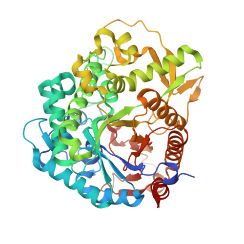
    3WBA, 3WBE

  • PubMed Abstract: 

    In order to identify a rice gibberellin ester β-D-glucosidase, gibberellin A4 β-D-glucosyl ester (GA4-GE) was synthesized and used to screen rice β-glucosidases. Os3BGlu6 was found to have the highest hydrolysis activity to GA4-GE among five recombinantly expressed rice glycoside hydrolase family GH1 enzymes from different phylogenic clusters. The kinetic parameters of Os3BGlu6 and its mutants E178Q, E178A, E394D, E394Q and M251N for hydrolysis of p-nitrophenyl β-D-glucopyranoside (pNPGlc) and GA4-GE confirmed the roles of the catalytic acid/base and nucleophile for hydrolysis of both substrates and suggested M251 contributes to binding hydrophobic aglycones. The activities of the Os3BGlu6 E178Q and E178A acid/base mutants were rescued by azide, which they transglucosylate to produce β-D-glucopyranosyl azide, in a pH-dependent manner, while acetate also rescued Os3BGlu6 E178A at low pH. High concentrations of sodium azide (200-400 mM) inhibited Os3BGlu6 E178Q but not Os3BGlu6 E178A. The structures of Os3BGlu6 E178Q crystallized with either GA4-GE or pNPGlc had a native α-D-glucosyl moiety covalently linked to the catalytic nucleophile, E394, which showed the hydrogen bonding to the 2-hydroxyl in the covalent intermediate. These data suggest that a GH1 β-glucosidase uses the same retaining catalytic mechanism to hydrolyze 1-O-acyl glucose ester and glucoside.


  • Organizational Affiliation

    School of Biochemistry and Chemistry, Institute of Science, Suranaree University of Technology, Nakhon Ratchasima, Thailand.


Macromolecules
Find similar proteins by:  (by identity cutoff)  |  3D Structure
Entity ID: 1
MoleculeChains Sequence LengthOrganismDetailsImage
Beta-glucosidase 6489Oryza sativa Japonica GroupMutation(s): 1 
Gene Names: BGLU6LOC_Os03g11420Os03g0212800
EC: 3.2.1.21
UniProt
Find proteins for Q8L7J2 (Oryza sativa subsp. japonica)
Explore Q8L7J2 
Go to UniProtKB:  Q8L7J2
Entity Groups  
Sequence Clusters30% Identity50% Identity70% Identity90% Identity95% Identity100% Identity
UniProt GroupQ8L7J2
Sequence Annotations
Expand
  • Reference Sequence
Experimental Data & Validation

Experimental Data

  • Method: X-RAY DIFFRACTION
  • Resolution: 1.90 Å
  • R-Value Free: 0.179 
  • R-Value Work: 0.146 
  • R-Value Observed: 0.148 
  • Space Group: P 21 21 21
Unit Cell:
Length ( Å )Angle ( ˚ )
a = 56.986α = 90
b = 91.127β = 90
c = 111.221γ = 90
Software Package:
Software NamePurpose
HKL-2000data collection
REFMACrefinement
HKL-2000data reduction
HKL-2000data scaling
REFMACphasing

Structure Validation

View Full Validation Report


Ligand Structure Quality Assessment 


Entry History 

Deposition Data

Revision History  (Full details and data files)

  • Version 1.0: 2013-09-04
    Type: Initial release
  • Version 1.1: 2020-07-29
    Type: Remediation
    Reason: Carbohydrate remediation
    Changes: Data collection, Database references, Derived calculations, Structure summary
  • Version 1.2: 2023-11-08
    Changes: Data collection, Database references, Refinement description, Structure summary