3W6L

Contribution of disulfide bond toward thermostability in hyperthermostable endocellulase


Experimental Data Snapshot

  • Method: X-RAY DIFFRACTION
  • Resolution: 1.75 Å
  • R-Value Free: 0.205 
  • R-Value Work: 0.176 
  • R-Value Observed: 0.178 

wwPDB Validation   3D Report Full Report


This is version 1.2 of the entry. See complete history


Literature

The role of disulfide bond in hyperthermophilic endocellulase

Kim, H.-W.Ishikawa, K.

(2013) Extremophiles 17: 593-599

  • DOI: https://doi.org/10.1007/s00792-013-0542-8
  • Primary Citation of Related Structures:  
    3W6L, 3W6M

  • PubMed Abstract: 

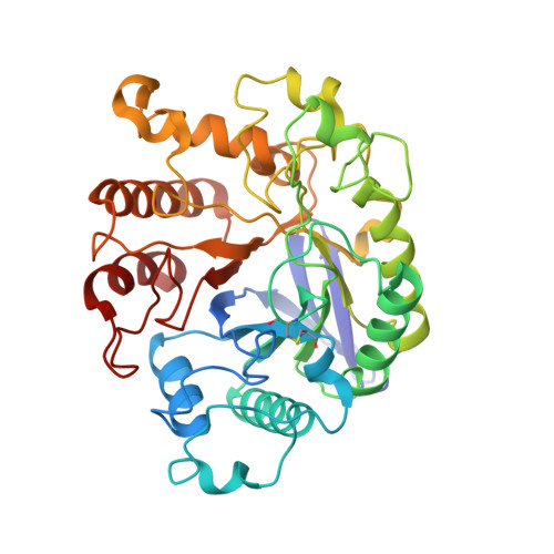
    The hyperthermophilic endocellulase, EGPh (glycosyl hydrolase family 5) from Pyrococcus horikoshii possesses 4 cysteine residues forming 2 disulfide bonds, as identified by structural analysis. One of the disulfide bonds is located at the proximal region of the active site in EGPh, which exhibits a distinct pattern from that of the thermophilic endocellulase EGAc (glycosyl hydrolase family 5) of Acidothermus cellulolyticus despite the structural similarity between the two endocellulases. The structural similarity between EGPh and EGAc suggests that EGPh possesses a structure suitable for changing the position of the disulfide bond corresponding to that in EGAc. Introduction of this alternative disulfide bond in EGPh, while removing the original disulfide bond, did not result in a loss of enzymatic activity but the EGPh was no longer hyperthermostable. These results suggest that the contribution of disulfide bond to hyperthermostability at temperature higher than 100 °C is restrictive, and that its impact is dependent on the specific structural environment of the hyperthermophilic proteins. The data suggest that the structural position and environment of the disulfide bond has a greater effect on high-temperature thermostability of the enzyme than on the potential energy of the dihedral angle that contributes to disulfide bond cleavage.


  • Organizational Affiliation

    Division of Life Sciences, Korea Polar Research Institute KOPRI, Incheon 406-840, Korea.


Macromolecules
Find similar proteins by:  (by identity cutoff)  |  3D Structure
Entity ID: 1
MoleculeChains Sequence LengthOrganismDetailsImage
458aa long hypothetical endo-1,4-beta-glucanase
A, B, C
377Pyrococcus horikoshii OT3Mutation(s): 0 
Gene Names: PH1171
EC: 3.2.1.4
UniProt
Find proteins for O58925 (Pyrococcus horikoshii (strain ATCC 700860 / DSM 12428 / JCM 9974 / NBRC 100139 / OT-3))
Explore O58925 
Go to UniProtKB:  O58925
Entity Groups  
Sequence Clusters30% Identity50% Identity70% Identity90% Identity95% Identity100% Identity
UniProt GroupO58925
Sequence Annotations
Expand
  • Reference Sequence
Small Molecules
Ligands 1 Unique
IDChains Name / Formula / InChI Key2D Diagram3D Interactions
PO4
Query on PO4

Download Ideal Coordinates CCD File 
D [auth A]
E [auth A]
F [auth A]
G [auth A]
H [auth B]
D [auth A],
E [auth A],
F [auth A],
G [auth A],
H [auth B],
I [auth B],
J [auth B],
K [auth B],
L [auth B],
M [auth B],
N [auth B],
O [auth B],
P [auth C],
Q [auth C],
R [auth C]
PHOSPHATE ION
O4 P
NBIIXXVUZAFLBC-UHFFFAOYSA-K
Experimental Data & Validation

Experimental Data

  • Method: X-RAY DIFFRACTION
  • Resolution: 1.75 Å
  • R-Value Free: 0.205 
  • R-Value Work: 0.176 
  • R-Value Observed: 0.178 
  • Space Group: C 1 2 1
Unit Cell:
Length ( Å )Angle ( ˚ )
a = 163.103α = 90
b = 58.34β = 109.65
c = 137.915γ = 90
Software Package:
Software NamePurpose
DENZOdata reduction
SCALEPACKdata scaling
PHENIXrefinement
PDB_EXTRACTdata extraction
HKL-2000data collection
HKL-2000data reduction

Structure Validation

View Full Validation Report


Entry History 

Deposition Data

Revision History  (Full details and data files)

  • Version 1.0: 2013-05-29
    Type: Initial release
  • Version 1.1: 2013-08-07
    Changes: Database references
  • Version 1.2: 2017-11-22
    Changes: Refinement description