3W0O

Crystal structure of a thermostable mutant of aminoglycoside phosphotransferase APH(4)-Ia, ternary complex with ADP and hygromycin B


Experimental Data Snapshot

  • Method: X-RAY DIFFRACTION
  • Resolution: 1.50 Å
  • R-Value Free: 0.212 
  • R-Value Work: 0.184 
  • R-Value Observed: 0.185 

wwPDB Validation   3D Report Full Report


Ligand Structure Quality Assessment 


This is version 1.1 of the entry. See complete history


Literature

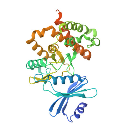
Crystal structures of the ternary complex of APH(4)-Ia/Hph with hygromycin B and an ATP analog using a thermostable mutant.

Iino, D.Takakura, Y.Fukano, K.Sasaki, Y.Hoshino, T.Ohsawa, K.Nakamura, A.Yajima, S.

(2013) J Struct Biol 183: 76-85

  • DOI: https://doi.org/10.1016/j.jsb.2013.05.023
  • Primary Citation of Related Structures:  
    3W0M, 3W0N, 3W0O, 3W0P, 3W0Q, 3W0R, 3W0S

  • PubMed Abstract: 

    Aminoglycoside 4-phosphotransferase-Ia (APH(4)-Ia)/Hygromycin B phosphotransferase (Hph) inactivates the aminoglycoside antibiotic hygromycin B (hygB) via phosphorylation. The crystal structure of the binary complex of APH(4)-Ia with hygB was recently reported. To characterize substrate recognition by the enzyme, we determined the crystal structure of the ternary complex of non-hydrolyzable ATP analog AMP-PNP and hygB with wild-type, thermostable Hph mutant Hph5, and apo-mutant enzyme forms. The comparison between the ternary complex and apo structures revealed that Hph undergoes domain movement upon binding of AMP-PNP and hygB. This was about half amount of the case of APH(9)-Ia. We also determined the crystal structures of mutants in which the conserved, catalytically important residues Asp198 and Asn203, and the non-conserved Asn202, were converted to Ala, revealing the importance of Asn202 for catalysis. Hph5 contains five amino acid substitutions that alter its thermostability by 16°C; its structure revealed that 4/5 mutations in Hph5 are located in the hydrophobic core and appear to increase thermostability by strengthening hydrophobic interactions.


  • Organizational Affiliation

    Department of Bioscience, Tokyo University of Agriculture, 1-1-1 Sakuragaoka, Setagaya-ku, Tokyo 156-8502, Japan.


Macromolecules
Find similar proteins by:  (by identity cutoff)  |  3D Structure
Entity ID: 1
MoleculeChains Sequence LengthOrganismDetailsImage
Hygromycin-B 4-O-kinase349Escherichia coliMutation(s): 5 
Gene Names: hph
EC: 2.7.1.163
UniProt
Find proteins for P00557 (Escherichia coli)
Explore P00557 
Go to UniProtKB:  P00557
Entity Groups  
Sequence Clusters30% Identity50% Identity70% Identity90% Identity95% Identity100% Identity
UniProt GroupP00557
Sequence Annotations
Expand
  • Reference Sequence
Small Molecules
Ligands 2 Unique
IDChains Name / Formula / InChI Key2D Diagram3D Interactions
HY0
Query on HY0

Download Ideal Coordinates CCD File 
C [auth A]HYGROMYCIN B VARIANT
C20 H37 N3 O13
GRRNUXAQVGOGFE-XFOBNZBXSA-N
ADP
Query on ADP

Download Ideal Coordinates CCD File 
B [auth A]ADENOSINE-5'-DIPHOSPHATE
C10 H15 N5 O10 P2
XTWYTFMLZFPYCI-KQYNXXCUSA-N
Experimental Data & Validation

Experimental Data

  • Method: X-RAY DIFFRACTION
  • Resolution: 1.50 Å
  • R-Value Free: 0.212 
  • R-Value Work: 0.184 
  • R-Value Observed: 0.185 
  • Space Group: P 32 2 1
Unit Cell:
Length ( Å )Angle ( ˚ )
a = 70.127α = 90
b = 70.127β = 90
c = 123.865γ = 120
Software Package:
Software NamePurpose
ADSCdata collection
MOLREPphasing
REFMACrefinement
MOSFLMdata reduction
SCALAdata scaling

Structure Validation

View Full Validation Report


Ligand Structure Quality Assessment 


Entry History 

Deposition Data

Revision History  (Full details and data files)

  • Version 1.0: 2013-08-07
    Type: Initial release
  • Version 1.1: 2024-03-20
    Changes: Data collection, Database references, Derived calculations, Structure summary