3VU1

Crystal structure of the C-terminal globular domain of oligosaccharyltransferase (PhAglB-L, O74088_PYRHO) from Pyrococcus horikoshii


Experimental Data Snapshot

  • Method: X-RAY DIFFRACTION
  • Resolution: 2.70 Å
  • R-Value Free: 0.215 
  • R-Value Work: 0.171 
  • R-Value Observed: 0.173 

wwPDB Validation   3D Report Full Report


This is version 1.1 of the entry. See complete history


Literature

Crystallographic and NMR Evidence for Flexibility in Oligosaccharyltransferases and Its Catalytic Significance

Nyirenda, J.Matsumoto, S.Saitoh, T.Maita, N.Noda, N.N.Inagaki, F.Kohda, D.

(2013) Structure 21: 32-41

  • DOI: https://doi.org/10.1016/j.str.2012.10.011
  • Primary Citation of Related Structures:  
    3VU0, 3VU1

  • PubMed Abstract: 

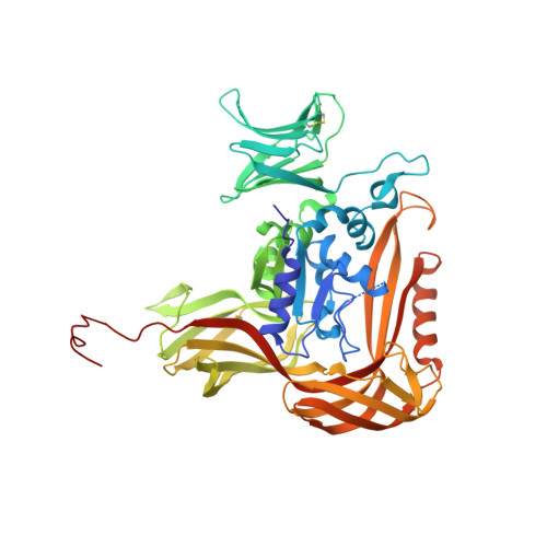
    Oligosaccharyltransferase (OST) is a membrane-bound enzyme that catalyzes the transfer of an oligosaccharide to an asparagine residue in glycoproteins. It possesses a binding pocket that recognizes Ser and Thr residues at the +2 position in the N-glycosylation consensus, Asn-X-Ser/Thr. We determined the crystal structures of the C-terminal globular domains of the catalytic subunits of two archaeal OSTs. A comparison with previously determined structures identified a segment with remarkable conformational plasticity, induced by crystal contact effects. We characterized its dynamic properties in solution by (15)N NMR relaxation analyses. Intriguingly, the mobile region contains the +2 Ser/Thr-binding pocket. In agreement, the flexibility restriction forced by an engineered disulfide crosslink abolished the enzymatic activity, and its cleavage fully restored activity. These results suggest the necessity of multiple conformational states in the reaction. The dynamic nature of the Ser/Thr pocket could facilitate the efficient scanning of N-glycosylation sequons along nascent polypeptide chains.


  • Organizational Affiliation

    Division of Structural Biology, Medical Institute of Bioregulation, Kyushu University, Fukuoka 812-8582, Japan.


Macromolecules
Find similar proteins by:  (by identity cutoff)  |  3D Structure
Entity ID: 1
MoleculeChains Sequence LengthOrganismDetailsImage
Putative uncharacterized protein PH0242
A, B
506Pyrococcus horikoshii OT3Mutation(s): 0 
Gene Names: AGLB
EC: 2.4.1.119
UniProt
Find proteins for O74088 (Pyrococcus horikoshii (strain ATCC 700860 / DSM 12428 / JCM 9974 / NBRC 100139 / OT-3))
Explore O74088 
Go to UniProtKB:  O74088
Entity Groups  
Sequence Clusters30% Identity50% Identity70% Identity90% Identity95% Identity100% Identity
UniProt GroupO74088
Sequence Annotations
Expand
  • Reference Sequence
Experimental Data & Validation

Experimental Data

  • Method: X-RAY DIFFRACTION
  • Resolution: 2.70 Å
  • R-Value Free: 0.215 
  • R-Value Work: 0.171 
  • R-Value Observed: 0.173 
  • Space Group: P 21 21 21
Unit Cell:
Length ( Å )Angle ( ˚ )
a = 83.467α = 90
b = 94.844β = 90
c = 186.345γ = 90
Software Package:
Software NamePurpose
HKL-2000data collection
MOLREPphasing
REFMACrefinement
HKL-2000data reduction
HKL-2000data scaling

Structure Validation

View Full Validation Report


Entry History 

Deposition Data

Revision History  (Full details and data files)

  • Version 1.0: 2013-01-23
    Type: Initial release
  • Version 1.1: 2023-11-08
    Changes: Data collection, Database references, Derived calculations, Refinement description