3VJR

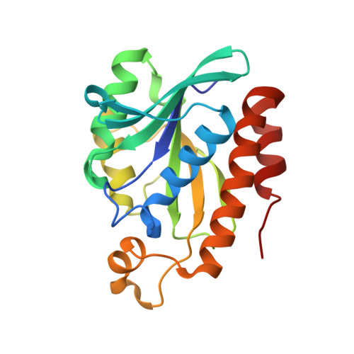
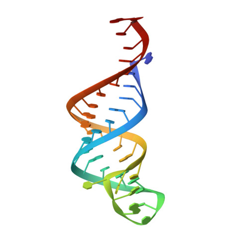
Crystal structure of Peptidyl-tRNA hydrolase from Escherichia coli in complex with the CCA-acceptor-T[PSI]C domain of tRNA


Experimental Data Snapshot

  • Method: X-RAY DIFFRACTION
  • Resolution: 2.40 Å
  • R-Value Free: 0.237 
  • R-Value Work: 0.193 
  • R-Value Observed: 0.195 

wwPDB Validation   3D Report Full Report


This is version 1.2 of the entry. See complete history


Literature

Structural basis for the substrate recognition and catalysis of peptidyl-tRNA hydrolase.

Ito, K.Murakami, R.Mochizuki, M.Qi, H.Shimizu, Y.Miura, K.Ueda, T.Uchiumi, T.

(2012) Nucleic Acids Res 40: 10521-10531

  • DOI: https://doi.org/10.1093/nar/gks790
  • Primary Citation of Related Structures:  
    3VJR

  • PubMed Abstract: 

    Peptidyl-tRNA hydrolase (Pth) cleaves the ester bond between the peptide and the tRNA of peptidyl-tRNA molecules, which are produced by aborted translation, to recycle tRNA for further rounds of protein synthesis. Pth is ubiquitous in nature, and its enzymatic activity is essential for bacterial viability. We have determined the crystal structure of Escherichia coli Pth in complex with the tRNA CCA-acceptor-TΨC domain, the enzyme-binding region of the tRNA moiety of the substrate, at 2.4 Å resolution. In combination with site-directed mutagenesis studies, the structure identified the amino acid residues involved in tRNA recognition. The structure also revealed that Pth interacts with the tRNA moiety through the backbone phosphates and riboses, and no base-specific interactions were observed, except for the interaction with the highly conserved base G53. This feature enables Pth to accept the diverse sequences of the elongator-tRNAs as substrate components. Furthermore, we propose an authentic Pth:peptidyl-tRNA complex model and a detailed mechanism for the hydrolysis reaction, based on the present crystal structure and the previous studies' results.


  • Organizational Affiliation

    Department of Biology, Faculty of Science, Niigata University, 8050 Ikarashi 2-no-cho, Nishi-ku, Niigata 950-2181, Japan. k-ito@bio.sc.niigata-u.ac.jp


Macromolecules

Find similar proteins by:  (by identity cutoff)  |  3D Structure
Entity ID: 1
MoleculeChains Sequence LengthOrganismDetailsImage
Peptidyl-tRNA hydrolase
A, C
197Escherichia coli K-12Mutation(s): 0 
Gene Names: pthb1204JW1195
EC: 3.1.1.29
UniProt
Find proteins for P0A7D1 (Escherichia coli (strain K12))
Explore P0A7D1 
Go to UniProtKB:  P0A7D1
Entity Groups  
Sequence Clusters30% Identity50% Identity70% Identity90% Identity95% Identity100% Identity
UniProt GroupP0A7D1
Sequence Annotations
Expand
  • Reference Sequence
Find similar nucleic acids by:  (by identity cutoff)  |  3D Structure
Entity ID: 2
MoleculeChains LengthOrganismImage
tRNA CCA-acceptor
B, D
36N/A
Sequence Annotations
Expand
  • Reference Sequence
Experimental Data & Validation

Experimental Data

  • Method: X-RAY DIFFRACTION
  • Resolution: 2.40 Å
  • R-Value Free: 0.237 
  • R-Value Work: 0.193 
  • R-Value Observed: 0.195 
  • Space Group: P 61
Unit Cell:
Length ( Å )Angle ( ˚ )
a = 55.07α = 90
b = 55.07β = 90
c = 413.1γ = 120
Software Package:
Software NamePurpose
XDSdata scaling
MOLREPphasing
REFMACrefinement
XDSdata reduction

Structure Validation

View Full Validation Report


Entry History 

Deposition Data

Revision History  (Full details and data files)

  • Version 1.0: 2012-09-12
    Type: Initial release
  • Version 1.1: 2013-06-26
    Changes: Database references
  • Version 1.2: 2023-11-08
    Changes: Data collection, Database references, Refinement description