3VEH

Structure of a M. tuberculosis salicylate synthase, MbtI, in complex with an inhibitor methylAMT


Experimental Data Snapshot

  • Method: X-RAY DIFFRACTION
  • Resolution: 2.00 Å
  • R-Value Free: 0.236 
  • R-Value Work: 0.175 
  • R-Value Observed: 0.178 

wwPDB Validation   3D Report Full Report


Ligand Structure Quality Assessment 


This is version 1.3 of the entry. See complete history


Literature

Implications of binding mode and active site flexibility for inhibitor potency against the salicylate synthase from Mycobacterium tuberculosis.

Chi, G.Manos-Turvey, A.O'Connor, P.D.Johnston, J.M.Evans, G.L.Baker, E.N.Payne, R.J.Lott, J.S.Bulloch, E.M.

(2012) Biochemistry 51: 4868-4879

  • DOI: https://doi.org/10.1021/bi3002067
  • Primary Citation of Related Structures:  
    3RV6, 3RV7, 3RV8, 3RV9, 3ST6, 3VEH

  • PubMed Abstract: 

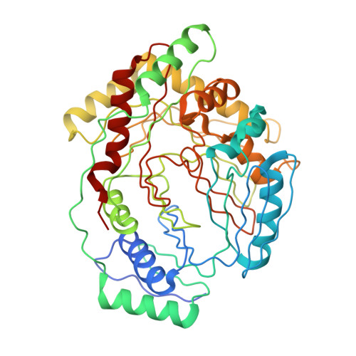
    MbtI is the salicylate synthase that catalyzes the first committed step in the synthesis of the iron chelating compound mycobactin in Mycobacterium tuberculosis. We previously developed a series of aromatic inhibitors against MbtI based on the reaction intermediate for this enzyme, isochorismate. The most potent of these inhibitors had hydrophobic substituents, ranging in size from a methyl to a phenyl group, appended to the terminal alkene of the enolpyruvyl group. These compounds exhibited low micromolar inhibition constants against MbtI and were at least an order of magnitude more potent than the parental compound for the series, which carries a native enolpyruvyl group. In this study, we sought to understand how the substituted enolpyruvyl group confers greater potency, by determining cocrystal structures of MbtI with six inhibitors from the series. A switch in binding mode at the MbtI active site is observed for inhibitors carrying a substituted enolpyruvyl group, relative to the parental compound. Computational studies suggest that the change in binding mode, and higher potency, is due to the effect of the substituents on the conformational landscape of the core inhibitor structure. The crystal structures and fluorescence-based thermal shift assays indicate that substituents larger than a methyl group are accommodated in the MbtI active site through significant but localized flexibility in the peptide backbone. These findings have implications for the design of improved inhibitors of MbtI, as well as other chorismate-utilizing enzymes from this family.


  • Organizational Affiliation

    School of Biological Sciences and Maurice Wilkins Centre for Molecular Biodiscovery, The University of Auckland, 3 Symonds Street, Private Bag 92019, Auckland 1142, New Zealand.


Macromolecules
Find similar proteins by:  (by identity cutoff)  |  3D Structure
Entity ID: 1
MoleculeChains Sequence LengthOrganismDetailsImage
Isochorismate synthase/isochorismate-pyruvate lyase mbtI
A, B, C, D
451Mycobacterium tuberculosisMutation(s): 0 
Gene Names: mbtItrpE2Rv2386cMT2454
EC: 4.1.3 (PDB Primary Data), 5.4.4.2 (PDB Primary Data)
UniProt
Find proteins for P9WFX1 (Mycobacterium tuberculosis (strain ATCC 25618 / H37Rv))
Explore P9WFX1 
Go to UniProtKB:  P9WFX1
Entity Groups  
Sequence Clusters30% Identity50% Identity70% Identity90% Identity95% Identity100% Identity
UniProt GroupP9WFX1
Sequence Annotations
Expand
  • Reference Sequence
Small Molecules
Ligands 5 Unique
IDChains Name / Formula / InChI Key2D Diagram3D Interactions
0GA
Query on 0GA

Download Ideal Coordinates CCD File 
E [auth A],
H [auth B],
L [auth C],
R [auth D]
3-{[(1Z)-1-carboxyprop-1-en-1-yl]oxy}-2-hydroxybenzoic acid
C11 H10 O6
MPHSNGOSXHODDD-UQCOIBPSSA-N
PEG
Query on PEG

Download Ideal Coordinates CCD File 
N [auth C],
P [auth C]
DI(HYDROXYETHYL)ETHER
C4 H10 O3
MTHSVFCYNBDYFN-UHFFFAOYSA-N
GOL
Query on GOL

Download Ideal Coordinates CCD File 
G [auth A],
J [auth B],
O [auth C],
Q [auth C]
GLYCEROL
C3 H8 O3
PEDCQBHIVMGVHV-UHFFFAOYSA-N
SCN
Query on SCN

Download Ideal Coordinates CCD File 
F [auth A],
I [auth B],
M [auth C]
THIOCYANATE ION
C N S
ZMZDMBWJUHKJPS-UHFFFAOYSA-M
K
Query on K

Download Ideal Coordinates CCD File 
K [auth B],
S [auth D]
POTASSIUM ION
K
NPYPAHLBTDXSSS-UHFFFAOYSA-N
Binding Affinity Annotations 
IDSourceBinding Affinity
0GA Binding MOAD:  3VEH Ki: 1.10e+4 (nM) from 1 assay(s)
PDBBind:  3VEH Ki: 1.10e+4 (nM) from 1 assay(s)
Experimental Data & Validation

Experimental Data

  • Method: X-RAY DIFFRACTION
  • Resolution: 2.00 Å
  • R-Value Free: 0.236 
  • R-Value Work: 0.175 
  • R-Value Observed: 0.178 
  • Space Group: P 1 21 1
Unit Cell:
Length ( Å )Angle ( ˚ )
a = 87.674α = 90
b = 115.955β = 91.59
c = 94.715γ = 90
Software Package:
Software NamePurpose
Blu-Icedata collection
MOLREPphasing
REFMACrefinement
HKL-2000data reduction
SCALEPACKdata scaling

Structure Validation

View Full Validation Report


Ligand Structure Quality Assessment 


Entry History 

Revision History  (Full details and data files)

  • Version 1.0: 2012-06-13
    Type: Initial release
  • Version 1.1: 2013-01-09
    Changes: Database references
  • Version 1.2: 2014-02-12
    Changes: Non-polymer description
  • Version 1.3: 2024-02-28
    Changes: Data collection, Database references, Derived calculations