3VA8

Crystal structure of enolase FG03645.1 (target EFI-502278) from Gibberella zeae PH-1 complexed with magnesium, formate and sulfate


Experimental Data Snapshot

  • Method: X-RAY DIFFRACTION
  • Resolution: 2.00 Å
  • R-Value Free: 0.217 
  • R-Value Work: 0.170 
  • R-Value Observed: 0.173 

wwPDB Validation   3D Report Full Report


This is version 1.2 of the entry. See complete history


Literature

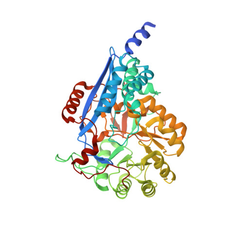
Crystal structure of dehydratase FG03645.1 from Gibberella zeae PH-1

Patskovsky, Y.Toro, R.Bhosle, R.Hillerich, B.Seidel, R.D.Washington, E.Scott Glenn, A.Chowdhury, S.Evans, B.Hammonds, J.Zencheck, W.D.Imker, H.J.Gerlt, J.A.Almo, S.C.

To be published.

Macromolecules
Find similar proteins by:  (by identity cutoff)  |  3D Structure
Entity ID: 1
MoleculeChains Sequence LengthOrganismDetailsImage
PROBABLE DEHYDRATASE445Fusarium graminearumMutation(s): 0 
Entity Groups  
Sequence Clusters30% Identity50% Identity70% Identity90% Identity95% Identity100% Identity
Sequence Annotations
Expand
  • Reference Sequence
Experimental Data & Validation

Experimental Data

  • Method: X-RAY DIFFRACTION
  • Resolution: 2.00 Å
  • R-Value Free: 0.217 
  • R-Value Work: 0.170 
  • R-Value Observed: 0.173 
  • Space Group: H 3 2
Unit Cell:
Length ( Å )Angle ( ˚ )
a = 135.669α = 90
b = 135.669β = 90
c = 164.246γ = 120
Software Package:
Software NamePurpose
PHASERphasing
REFMACrefinement
HKL-3000data reduction
HKL-3000data scaling

Structure Validation

View Full Validation Report


Entry History 

Revision History  (Full details and data files)

  • Version 1.0: 2012-02-01
    Type: Initial release
  • Version 1.1: 2012-03-07
    Changes: Structure summary
  • Version 1.2: 2023-09-13
    Changes: Data collection, Database references, Derived calculations, Refinement description