3V9R

Crystal structure of Saccharomyces cerevisiae MHF complex


Experimental Data Snapshot

  • Method: X-RAY DIFFRACTION
  • Resolution: 2.40 Å
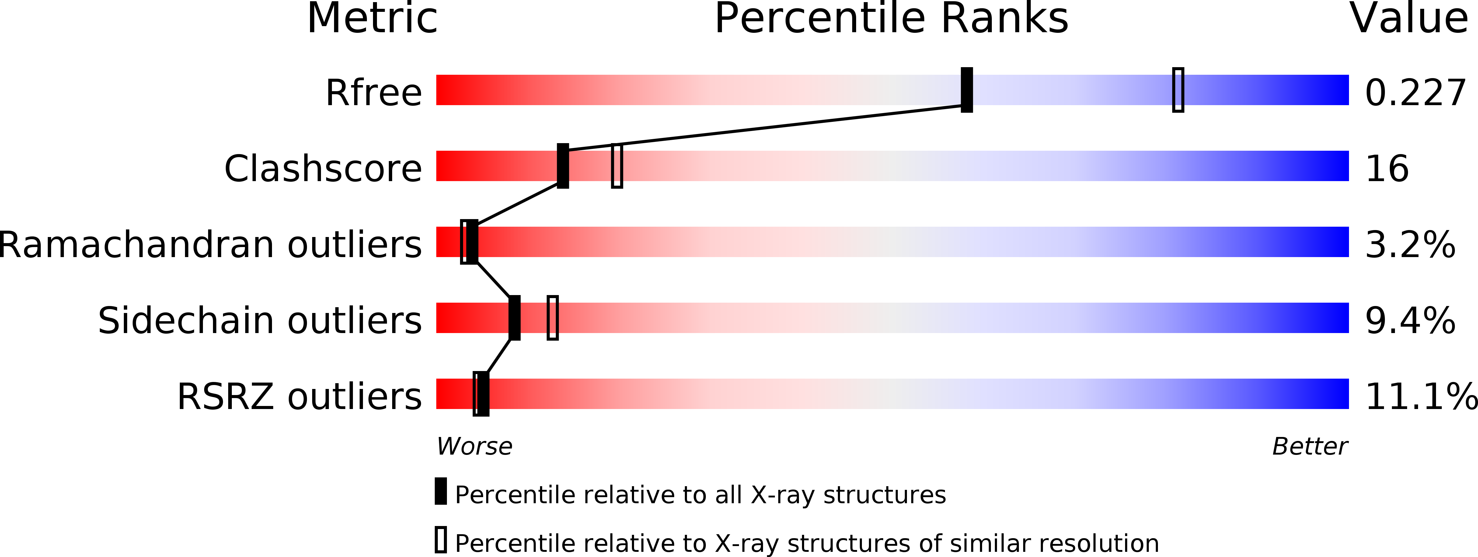
  • R-Value Free: 0.290 
  • R-Value Work: 0.239 
  • R-Value Observed: 0.242 

wwPDB Validation   3D Report Full Report


This is version 1.0 of the entry. See complete history


Literature

Saccharomyces Cerevisiae MHF Complex Structurally Resembles the Histones (H3-H4)(2) Heterotetramer and Functions as a Heterotetramer

Yang, H.Zhang, T.Tao, Y.Wu, L.Li, H.T.Zhou, J.Q.Zhong, C.Ding, J.

(2012) Structure 20: 364-370

  • DOI: https://doi.org/10.1016/j.str.2011.12.012
  • Primary Citation of Related Structures:  
    3V9R

  • PubMed Abstract: 

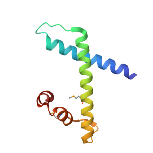
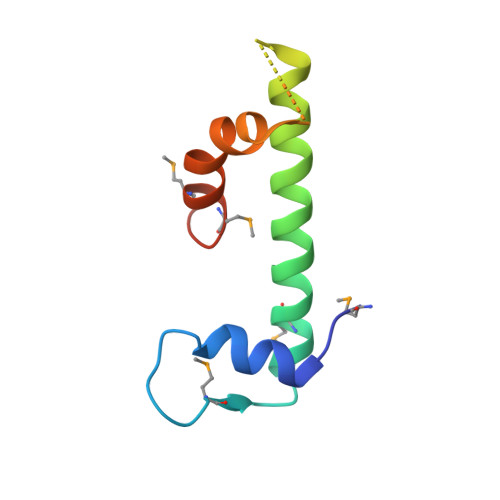
    Fanconi anemia (FA) is a chromosomal instability disorder associated with deficiencies in the Fanconi anemia complementation group (FANC) network. A complex consisting of FANCM-associated histone-fold proteins 1 and 2 (MHF1 and MHF2) has been shown to act cooperatively with FANCM in DNA damage repair in the FA pathway. Here we report the structure of Saccharomyces cerevisiae MHF complex in which MHF1 and MHF2 assume a typical histone fold, and the complex has a heterotetrameric architecture similar to that of the histones (H3-H4)₂ heterotetramer. Loop L2 of MHF1 is probably involved in DNA binding, and loop L3 and helices α2 and α3 of one MHF1 subunit interact with those of the other to form two heterotetramer interfaces. Further genetic data demonstrate that the heterotetramer assembly is essential for the function of the complex in DNA repair. These results provide, to the best of our knowledge, new mechanistic insights into the function of the MHF complex.


  • Organizational Affiliation

    State Key Laboratory of Molecular Biology and Research Center for Structural Biology, Institute of Biochemistry and Cell Biology, Shanghai Institutes for Biological Sciences, Chinese Academy of Sciences, 320 Yue Yang Road, Shanghai 200031, China.


Macromolecules
Find similar proteins by:  (by identity cutoff)  |  3D Structure
Entity ID: 1
MoleculeChains Sequence LengthOrganismDetailsImage
Uncharacterized protein YOL086W-A
A, C
90Saccharomyces cerevisiae S288CMutation(s): 0 
Gene Names: YOL086W-A
UniProt
Find proteins for Q3E835 (Saccharomyces cerevisiae (strain ATCC 204508 / S288c))
Explore Q3E835 
Go to UniProtKB:  Q3E835
Entity Groups  
Sequence Clusters30% Identity50% Identity70% Identity90% Identity95% Identity100% Identity
UniProt GroupQ3E835
Sequence Annotations
Expand
  • Reference Sequence
Find similar proteins by:  (by identity cutoff)  |  3D Structure
Entity ID: 2
MoleculeChains Sequence LengthOrganismDetailsImage
Uncharacterized protein YDL160C-A
B, D
88Saccharomyces cerevisiae S288CMutation(s): 0 
Gene Names: YDL160C-A
UniProt
Find proteins for Q3E829 (Saccharomyces cerevisiae (strain ATCC 204508 / S288c))
Explore Q3E829 
Go to UniProtKB:  Q3E829
Entity Groups  
Sequence Clusters30% Identity50% Identity70% Identity90% Identity95% Identity100% Identity
UniProt GroupQ3E829
Sequence Annotations
Expand
  • Reference Sequence
Small Molecules
Ligands 1 Unique
IDChains Name / Formula / InChI Key2D Diagram3D Interactions
SO4
Query on SO4

Download Ideal Coordinates CCD File 
E [auth A]SULFATE ION
O4 S
QAOWNCQODCNURD-UHFFFAOYSA-L
Modified Residues  1 Unique
IDChains TypeFormula2D DiagramParent
MSE
Query on MSE
A, C
L-PEPTIDE LINKINGC5 H11 N O2 SeMET
Experimental Data & Validation

Experimental Data

  • Method: X-RAY DIFFRACTION
  • Resolution: 2.40 Å
  • R-Value Free: 0.290 
  • R-Value Work: 0.239 
  • R-Value Observed: 0.242 
  • Space Group: C 1 2 1
Unit Cell:
Length ( Å )Angle ( ˚ )
a = 68.823α = 90
b = 104.065β = 109.09
c = 70.862γ = 90
Software Package:
Software NamePurpose
REFMACrefinement
PDB_EXTRACTdata extraction
HKL-2000data collection
HKL-2000data reduction
HKL-2000data scaling
AutoSolphasing

Structure Validation

View Full Validation Report


Entry History 

Deposition Data

Revision History  (Full details and data files)

  • Version 1.0: 2012-02-29
    Type: Initial release