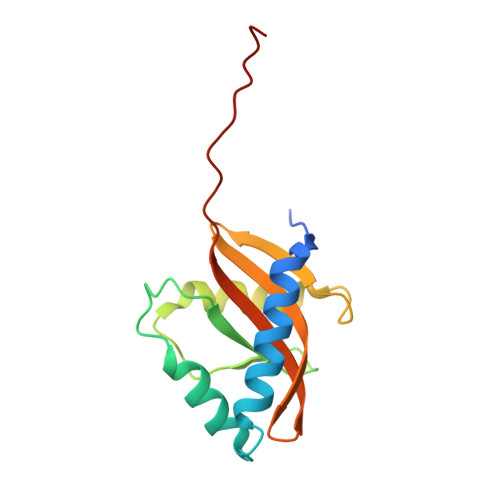
The crystal structure of the periplasmic domain of Vibrio parahaemolyticus CpxA
Kwon, E., Kim, D.Y., Ngo, T.D., Gross, C.A., Gross, J.D., Kim, K.K.(2012) Protein Sci 21: 1334-1343
- PubMed: 22760860
- DOI: https://doi.org/10.1002/pro.2120
- Primary Citation of Related Structures:
3V67 - PubMed Abstract:
The Cpx two-component system of Gram-negative bacteria senses extracytoplasmic stresses using the histidine kinase CpxA, a membrane-bound sensor, and controls the transcription of the genes involved in stress response by the cytosolic response regulator CpxR, which is activated by the phosphorelay from CpxA. CpxP, a CpxA-associated protein, also plays an important role in the regulation of the Cpx system by inhibiting the autophosphorylation of CpxA. Although the stress signals and physiological roles of the Cpx system have been extensively studied, the lack of structural information has limited the understanding of the detailed mechanism of ligand binding and regulation of CpxA. In this study, we solved the crystal structure of the periplasmic domain of Vibrio parahaemolyticus CpxA (VpCpxA-peri) to a resolution of 2.1 Å and investigated its interaction with CpxP. VpCpxA-peri has a globular Per-ARNT-SIM (PAS) domain and a protruded C-terminal tail, which may be required for ligand sensing and CpxP binding, respectively. The direct interaction of the PAS core of VpCpxA-peri with VpCpxP was not detected by NMR, suggesting that the C-terminal tail or other factors, such as the membrane environment, are necessary for the binding of CpxA to CpxP.
Organizational Affiliation:
Department of Molecular Cell Biology, Samsung Biomedical Research Institute, Sungkyunkwan University School of Medicine, Suwon 440-746, Korea.


