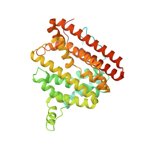
3V1V

Crystal structure of 2-methylisoborneol synthase from Streptomyces coelicolor A3(2) in complex with Mg2+ and geranyl-S-thiolodiphosphate


Experimental Data Snapshot

  • Method: X-RAY DIFFRACTION
  • Resolution: 1.80 Å
  • R-Value Free: 0.176 
  • R-Value Work: 0.150 
  • R-Value Observed: 0.151 

wwPDB Validation   3D Report Full Report


Ligand Structure Quality Assessment 


This is version 1.2 of the entry. See complete history


Literature

Structure of 2-Methylisoborneol Synthase from Streptomyces coelicolor and Implications for the Cyclization of a Noncanonical C-Methylated Monoterpenoid Substrate.

Koksal, M.Chou, W.K.Cane, D.E.Christianson, D.W.

(2012) Biochemistry 51: 3011-3020

  • DOI: https://doi.org/10.1021/bi201827a
  • Primary Citation of Related Structures:  
    3V1V, 3V1X

  • PubMed Abstract: 

    The crystal structure of 2-methylisoborneol synthase (MIBS) from Streptomyces coelicolor A3(2) has been determined in complex with substrate analogues geranyl-S-thiolodiphosphate and 2-fluorogeranyl diphosphate at 1.80 and 1.95 Å resolution, respectively. This terpenoid cyclase catalyzes the cyclization of the naturally occurring, noncanonical C-methylated isoprenoid substrate, 2-methylgeranyl diphosphate, to form the bicyclic product 2-methylisoborneol, a volatile C(11) homoterpene alcohol with an earthy, musty odor. While MIBS adopts the tertiary structure of a class I terpenoid cyclase, its dimeric quaternary structure differs from that previously observed in dimeric terpenoid cyclases from plants and fungi. The quaternary structure of MIBS is nonetheless similar in some respects to that of dimeric farnesyl diphosphate synthase, which is not a cyclase. The structures of MIBS complexed with substrate analogues provide insights regarding differences in the catalytic mechanism of MIBS and the mechanisms of (+)-bornyl diphosphate synthase and endo-fenchol synthase, plant cyclases that convert geranyl diphosphate into products with closely related bicyclic bornyl skeletons, but distinct structures and stereochemistries.


  • Organizational Affiliation

    Roy and Diana Vagelos Laboratories, Department of Chemistry, University of Pennsylvania, 231 South 34th Street, Philadelphia, Pennsylvania 19104-6323, USA.


Macromolecules
Find similar proteins by:  (by identity cutoff)  |  3D Structure
Entity ID: 1
MoleculeChains Sequence LengthOrganismDetailsImage
2-methylisoborneol synthase433Streptomyces coelicolor A3(2)Mutation(s): 0 
Gene Names: SC1A4.08SCBAC12C8.01SCO7700
EC: 4.2.3
UniProt
Find proteins for Q9F1Y6 (Streptomyces coelicolor (strain ATCC BAA-471 / A3(2) / M145))
Explore Q9F1Y6 
Go to UniProtKB:  Q9F1Y6
Entity Groups  
Sequence Clusters30% Identity50% Identity70% Identity90% Identity95% Identity100% Identity
UniProt GroupQ9F1Y6
Sequence Annotations
Expand
  • Reference Sequence
Experimental Data & Validation

Experimental Data

  • Method: X-RAY DIFFRACTION
  • Resolution: 1.80 Å
  • R-Value Free: 0.176 
  • R-Value Work: 0.150 
  • R-Value Observed: 0.151 
  • Space Group: P 43 2 2
Unit Cell:
Length ( Å )Angle ( ˚ )
a = 99.392α = 90
b = 99.392β = 90
c = 105.742γ = 90
Software Package:
Software NamePurpose
CBASSdata collection
PHENIXmodel building
PHENIXrefinement
HKL-2000data reduction
HKL-2000data scaling
PHENIXphasing

Structure Validation

View Full Validation Report


Ligand Structure Quality Assessment 


Entry History 

Deposition Data

Revision History  (Full details and data files)

  • Version 1.0: 2012-04-11
    Type: Initial release
  • Version 1.1: 2012-04-25
    Changes: Database references
  • Version 1.2: 2024-02-28
    Changes: Data collection, Database references, Derived calculations