


Crystal structure of an oxidoreductase from Sinorhizobium meliloti 1021
Agarwal, R., Almo, S.C., Swaminathan, S.To be published.
Experimental Data Snapshot
wwPDB Validation 3D Report Full Report

Entity ID: 1 | |||||
|---|---|---|---|---|---|
| Molecule | Chains | Sequence Length | Organism | Details | Image |
| Probable oxidoreductase | 260 | Sinorhizobium meliloti | Mutation(s): 0 Gene Names: y20210, RB0203, SM_b20210 EC: 1.1.1.100 | 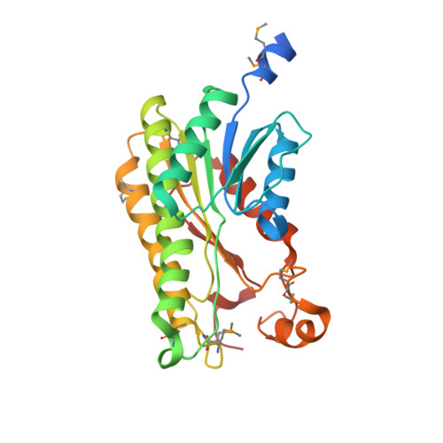 | |
UniProt | |||||
Find proteins for Q7ANT0 (Rhizobium meliloti (strain 1021)) Explore Q7ANT0 Go to UniProtKB: Q7ANT0 | |||||
Entity Groups | |||||
| Sequence Clusters | 30% Identity50% Identity70% Identity90% Identity95% Identity100% Identity | ||||
| UniProt Group | Q7ANT0 | ||||
Sequence AnnotationsExpand | |||||
| |||||
| Ligands 1 Unique | |||||
|---|---|---|---|---|---|
| ID | Chains | Name / Formula / InChI Key | 2D Diagram | 3D Interactions | |
| PO4 Query on PO4 | E [auth C], F [auth D] | PHOSPHATE ION O4 P NBIIXXVUZAFLBC-UHFFFAOYSA-K |  | ||
| Modified Residues 1 Unique | |||||
|---|---|---|---|---|---|
| ID | Chains | Type | Formula | 2D Diagram | Parent |
| MSE Query on MSE | A, B, C, D | L-PEPTIDE LINKING | C5 H11 N O2 Se |  | MET |
| Length ( Å ) | Angle ( ˚ ) |
|---|---|
| a = 84.225 | α = 90 |
| b = 84.225 | β = 90 |
| c = 249.238 | γ = 120 |
| Software Name | Purpose |
|---|---|
| REFMAC | refinement |
RCSB PDB (citation) is hosted by
RCSB PDB is a member of the

