3UAN

Crystal structure of 3-O-sulfotransferase (3-OST-1) with bound PAP and heptasaccharide substrate


Experimental Data Snapshot

  • Method: X-RAY DIFFRACTION
  • Resolution: 1.84 Å
  • R-Value Free: 0.202 
  • R-Value Work: 0.174 
  • R-Value Observed: 0.175 

wwPDB Validation   3D Report Full Report


Ligand Structure Quality Assessment 


This is version 2.1 of the entry. See complete history


Literature

Dissecting the substrate recognition of 3-O-sulfotransferase for the biosynthesis of anticoagulant heparin.

Moon, A.F.Xu, Y.Woody, S.M.Krahn, J.M.Linhardt, R.J.Liu, J.Pedersen, L.C.

(2012) Proc Natl Acad Sci U S A 109: 5265-5270

  • DOI: https://doi.org/10.1073/pnas.1117923109
  • Primary Citation of Related Structures:  
    3UAN

  • PubMed Abstract: 

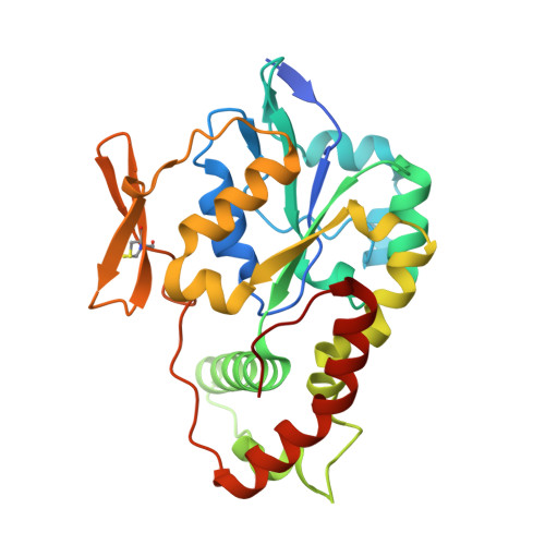
    Heparin is a polysaccharide-based natural product that is used clinically as an anticoagulant drug. Heparan sulfate 3-O-sulfotransferase (3-OST) is an enzyme that transfers a sulfo group to the 3-OH position of a glucosamine unit. 3-OST is present in multiple isoforms, and the polysaccharides modified by these different isoforms perform distinct biological functions. 3-OST isoform 1 (3-OST-1) is the key enzyme for the biosynthesis of anticoagulant heparin. Here, we report the crystal structure of the ternary complex of 3-OST-1, 3'-phosphoadenosine 5'-phosphate, and a heptasaccharide substrate. Comparisons to previously determined structures of 3-OST-3 reveal unique binding modes used by the different isoforms of 3-OST for distinguishing the fine structures of saccharide substrates. Our data demonstrate that the saccharide substrates display distinct conformations when interacting with the different 3-OST isoforms. Site-directed mutagenesis data suggest that several key amino residues, including Lys259, Thr256, and Trp283 in 3-OST-3 and Arg268 in 3-OST-1, play important roles in substrate binding and specificity between isoforms. These results deepen our understanding of the biosynthetic mechanism of heparan sulfate and provide structural information for engineering enzymes for an enhanced biosynthetic approach to heparin production.


  • Organizational Affiliation

    Laboratory of Structural Biology, National Institute of Environmental Health Sciences, National Institutes of Health, Research Triangle Park, NC 27709, USA.


Macromolecules
Find similar proteins by:  (by identity cutoff)  |  3D Structure
Entity ID: 1
MoleculeChains Sequence LengthOrganismDetailsImage
Heparan sulfate glucosamine 3-O-sulfotransferase 1
A, B
269Mus musculusMutation(s): 0 
Gene Names: Hs3st13ost3ost1
EC: 2.8.2.23
UniProt
Find proteins for O35310 (Mus musculus)
Explore O35310 
Go to UniProtKB:  O35310
Entity Groups  
Sequence Clusters30% Identity50% Identity70% Identity90% Identity95% Identity100% Identity
UniProt GroupO35310
Sequence Annotations
Expand
  • Reference Sequence
Oligosaccharides

Help

Entity ID: 2
MoleculeChains Length2D Diagram Glycosylation3D Interactions
2-acetamido-2-deoxy-6-O-sulfo-alpha-D-glucopyranose-(1-4)-beta-D-glucopyranuronic acid-(1-4)-2-deoxy-6-O-sulfo-2-(sulfoamino)-alpha-D-glucopyranose-(1-4)-2-O-sulfo-alpha-L-idopyranuronic acid-(1-4)-2-deoxy-6-O-sulfo-2-(sulfoamino)-alpha-D-glucopyranose-(1-4)-beta-D-glucopyranuronic acid
C
6N/A
Glycosylation Resources
GlyTouCan:  G04206TC
GlyCosmos:  G04206TC
Entity ID: 3
MoleculeChains Length2D Diagram Glycosylation3D Interactions
2-acetamido-2-deoxy-6-O-sulfo-alpha-D-glucopyranose-(1-4)-beta-D-glucopyranuronic acid-(1-4)-2-deoxy-6-O-sulfo-2-(sulfoamino)-alpha-D-glucopyranose-(1-4)-2-O-sulfo-alpha-L-idopyranuronic acid-(1-4)-2-deoxy-6-O-sulfo-2-(sulfoamino)-alpha-D-glucopyranose
D
5N/A
Glycosylation Resources
GlyTouCan:  G46176LD
GlyCosmos:  G46176LD
Experimental Data & Validation

Experimental Data

  • Method: X-RAY DIFFRACTION
  • Resolution: 1.84 Å
  • R-Value Free: 0.202 
  • R-Value Work: 0.174 
  • R-Value Observed: 0.175 
  • Space Group: P 1 21 1
Unit Cell:
Length ( Å )Angle ( ˚ )
a = 52.883α = 90
b = 62.168β = 95.16
c = 86.373γ = 90
Software Package:
Software NamePurpose
StructureStudiodata collection
MOLREPphasing
PHENIXrefinement
HKL-2000data reduction
HKL-2000data scaling

Structure Validation

View Full Validation Report


Ligand Structure Quality Assessment 


Entry History 

Deposition Data

Revision History  (Full details and data files)

  • Version 1.0: 2012-04-04
    Type: Initial release
  • Version 1.1: 2012-04-18
    Changes: Database references
  • Version 1.2: 2012-04-25
    Changes: Database references
  • Version 2.0: 2020-07-29
    Type: Remediation
    Reason: Carbohydrate remediation
    Changes: Advisory, Atomic model, Data collection, Database references, Derived calculations, Structure summary
  • Version 2.1: 2023-09-13
    Changes: Data collection, Database references, Refinement description, Structure summary