3U7G

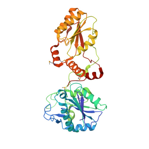
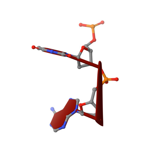
Crystal structure of mPNKP catalytic fragment (D170A) bound to single-stranded DNA (TCCTAp)


Experimental Data Snapshot

  • Method: X-RAY DIFFRACTION
  • Resolution: 2.10 Å
  • R-Value Free: 0.260 
  • R-Value Work: 0.207 
  • R-Value Observed: 0.210 

wwPDB Validation   3D Report Full Report


This is version 1.2 of the entry. See complete history


Literature

Structural basis for the phosphatase activity of polynucleotide kinase/phosphatase on single- and double-stranded DNA substrates.

Coquelle, N.Havali-Shahriari, Z.Bernstein, N.Green, R.Glover, J.N.

(2011) Proc Natl Acad Sci U S A 108: 21022-21027

  • DOI: https://doi.org/10.1073/pnas.1112036108
  • Primary Citation of Related Structures:  
    3U7E, 3U7F, 3U7G, 3U7H

  • PubMed Abstract: 

    Polynucleotide kinase/phosphatase (PNKP) is a critical mammalian DNA repair enzyme that generates 5'-phosphate and 3'-hydroxyl groups at damaged DNA termini that are required for subsequent processing by DNA ligases and polymerases. The PNKP phosphatase domain recognizes 3'-phosphate termini within DNA nicks, gaps, or at double- or single-strand breaks. Here we present a mechanistic rationale for the recognition of damaged DNA termini by the PNKP phosphatase domain. The crystal structures of PNKP bound to single-stranded DNA substrates reveals a narrow active site cleft that accommodates a single-stranded substrate in a sequence-independent manner. Biochemical studies suggest that the terminal base pairs of double-stranded substrates near the 3'-phosphate are destabilized by PNKP to allow substrate access to the active site. A positively charged surface distinct from the active site specifically facilitates interactions with double-stranded substrates, providing a complex DNA binding surface that enables the recognition of diverse substrates.


  • Organizational Affiliation

    Department of Biochemistry, University of Alberta, Edmonton, AB, Canada T6G 2H7.


Macromolecules

Find similar proteins by:  (by identity cutoff)  |  3D Structure
Entity ID: 1
MoleculeChains Sequence LengthOrganismDetailsImage
Bifunctional polynucleotide phosphatase/kinase379Mus musculusMutation(s): 1 
Gene Names: Pnkp
EC: 3.1.3.32 (PDB Primary Data), 2.7.1.78 (PDB Primary Data)
UniProt & NIH Common Fund Data Resources
Find proteins for Q9JLV6 (Mus musculus)
Explore Q9JLV6 
Go to UniProtKB:  Q9JLV6
IMPC:  MGI:1891698
Entity Groups  
Sequence Clusters30% Identity50% Identity70% Identity90% Identity95% Identity100% Identity
UniProt GroupQ9JLV6
Sequence Annotations
Expand
  • Reference Sequence

Find similar nucleic acids by:  Sequence   |   3D Structure  

Entity ID: 2
MoleculeChains LengthOrganismImage
DNAB [auth X]5N/A
Sequence Annotations
Expand
  • Reference Sequence
Experimental Data & Validation

Experimental Data

  • Method: X-RAY DIFFRACTION
  • Resolution: 2.10 Å
  • R-Value Free: 0.260 
  • R-Value Work: 0.207 
  • R-Value Observed: 0.210 
  • Space Group: C 1 2 1
Unit Cell:
Length ( Å )Angle ( ˚ )
a = 77.16α = 90
b = 43.03β = 107.12
c = 127.27γ = 90
Software Package:
Software NamePurpose
XSCALEdata scaling
PHENIXrefinement
PDB_EXTRACTdata extraction
Blu-Icedata collection
XDSdata reduction
PHASERphasing

Structure Validation

View Full Validation Report


Entry History 

Deposition Data

Revision History  (Full details and data files)

  • Version 1.0: 2011-12-14
    Type: Initial release
  • Version 1.1: 2012-08-29
    Changes: Database references
  • Version 1.2: 2017-11-08
    Changes: Refinement description