3U73

Crystal structure of stabilized human uPAR mutant in complex with ATF


Experimental Data Snapshot

  • Method: X-RAY DIFFRACTION
  • Resolution: 3.19 Å
  • R-Value Free: 0.258 
  • R-Value Work: 0.216 
  • R-Value Observed: 0.218 

wwPDB Validation   3D Report Full Report


This is version 2.0 of the entry. See complete history


Literature

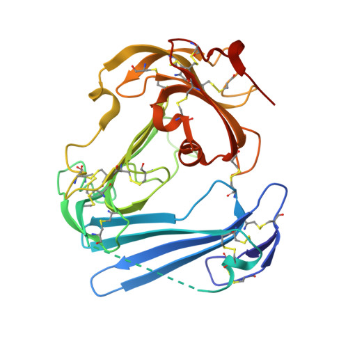
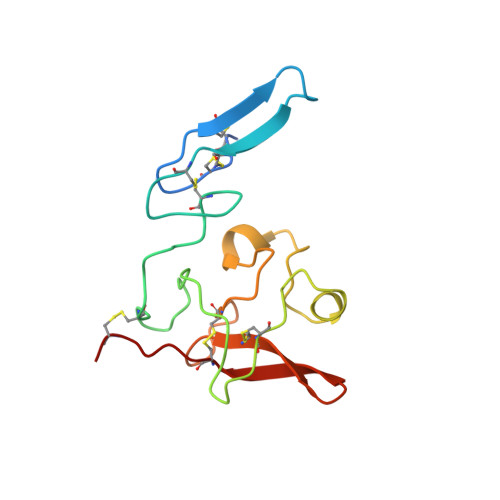
Crystal structure of the urokinase receptor in a ligand-free form.

Xu, X.Gardsvoll, H.Yuan, C.Lin, L.Ploug, M.Huang, M.

(2012) J Mol Biol 416: 629-641

  • DOI: https://doi.org/10.1016/j.jmb.2011.12.058
  • Primary Citation of Related Structures:  
    3U73, 3U74

  • PubMed Abstract: 

    The urokinase receptor urokinase-type plasminogen activator receptor (uPAR) is a surface receptor capable of not only focalizing urokinase-type plasminogen activator (uPA)-mediated fibrinolysis to the pericellular micro-environment but also promoting cell migration and chemotaxis. Consistent with this multifunctional role, uPAR binds several extracellular ligands, including uPA and vitronectin. Structural studies suggest that uPAR possesses structural flexibility. It is, however, not clear whether this flexibility is an inherent property of the uPAR structure per se or whether it is induced upon ligand binding. The crystal structure of human uPAR in its ligand-free state would clarify this issue, but such information remains unfortunately elusive. We now report the crystal structures of a stabilized, human uPAR (H47C/N259C) in its ligand-free form to 2.4 Å and in complex with amino-terminal fragment (ATF) to 3.2 Å. The structure of uPAR(H47C/N259C) in complex with ATF resembles the wild-type uPAR·ATF complex, demonstrating that these mutations do not perturb the uPA binding properties of uPAR. The present structure of uPAR(H47C/N259C) provides the first structural definition of uPAR in its ligand-free form, which represents one of the biologically active conformations of uPAR as defined by extensive biochemical studies. The domain boundary between uPAR DI-DII domains is more flexible than the DII-DIII domain boundary. Two important structural features are highlighted by the present uPAR structure. First, the DI-DIII domain boundary may face the cell membrane. Second, loop 130-140 of uPAR plays a dynamic role during ligand loading/unloading. Together, these studies provide new insights into uPAR structure-function relationships, emphasizing the importance of the inter-domain dynamics of this modular receptor.


  • Organizational Affiliation

    Beth Israel Deaconess Medical Center, Harvard Medical School, Boston, MA 02215, USA.


Macromolecules
Find similar proteins by:  (by identity cutoff)  |  3D Structure
Entity ID: 1
MoleculeChains Sequence LengthOrganismDetailsImage
Urokinase plasminogen activator surface receptorA [auth U]283Homo sapiensMutation(s): 2 
Gene Names: MO3PLAURUPAR
UniProt & NIH Common Fund Data Resources
Find proteins for Q03405 (Homo sapiens)
Explore Q03405 
Go to UniProtKB:  Q03405
PHAROS:  Q03405
GTEx:  ENSG00000011422 
Entity Groups  
Sequence Clusters30% Identity50% Identity70% Identity90% Identity95% Identity100% Identity
UniProt GroupQ03405
Sequence Annotations
Expand
  • Reference Sequence
Find similar proteins by:  (by identity cutoff)  |  3D Structure
Entity ID: 2
MoleculeChains Sequence LengthOrganismDetailsImage
Urokinase-type plasminogen activatorB [auth A]132Homo sapiensMutation(s): 0 
Gene Names: PLAU
EC: 3.4.21.73
UniProt & NIH Common Fund Data Resources
Find proteins for P00749 (Homo sapiens)
Explore P00749 
Go to UniProtKB:  P00749
PHAROS:  P00749
GTEx:  ENSG00000122861 
Entity Groups  
Sequence Clusters30% Identity50% Identity70% Identity90% Identity95% Identity100% Identity
UniProt GroupP00749
Sequence Annotations
Expand
  • Reference Sequence
Oligosaccharides

Help

Entity ID: 3
MoleculeChains Length2D Diagram Glycosylation3D Interactions
alpha-D-mannopyranose-(1-4)-2-acetamido-2-deoxy-beta-D-glucopyranose-(1-4)-2-acetamido-2-deoxy-beta-D-glucopyranoseC [auth B]3N-Glycosylation
Glycosylation Resources
GlyTouCan:  G62182OO
GlyCosmos:  G62182OO
GlyGen:  G62182OO
Entity ID: 4
MoleculeChains Length2D Diagram Glycosylation3D Interactions
2-acetamido-2-deoxy-beta-D-glucopyranose-(1-4)-2-acetamido-2-deoxy-beta-D-glucopyranoseD [auth C],
E [auth D]
2N-Glycosylation
Glycosylation Resources
GlyTouCan:  G42666HT
GlyCosmos:  G42666HT
GlyGen:  G42666HT
Experimental Data & Validation

Experimental Data

  • Method: X-RAY DIFFRACTION
  • Resolution: 3.19 Å
  • R-Value Free: 0.258 
  • R-Value Work: 0.216 
  • R-Value Observed: 0.218 
  • Space Group: P 31 2 1
Unit Cell:
Length ( Å )Angle ( ˚ )
a = 130.896α = 90
b = 130.896β = 90
c = 105.315γ = 120
Software Package:
Software NamePurpose
SCALEPACKdata scaling
REFMACrefinement
PDB_EXTRACTdata extraction

Structure Validation

View Full Validation Report


Entry History 

Deposition Data

Revision History  (Full details and data files)

  • Version 1.0: 2012-04-18
    Type: Initial release
  • Version 2.0: 2020-07-29
    Type: Remediation
    Reason: Carbohydrate remediation
    Changes: Advisory, Atomic model, Data collection, Database references, Derived calculations, Structure summary