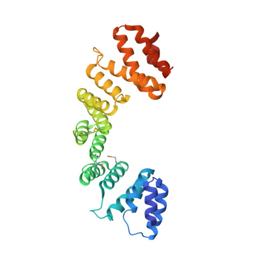
Crystal Structure of the C-terminal part of the TPR repeat-containing protein Q11TI6_CYTH3 from Cytophaga hutchinsonii.
Vorobiev, S., Neely, H., Chen, Y., Seetharaman, J., Patel, P., Xiao, R., Ciccosanti, C., Maglaqui, M., Everett, J.K., Nair, R., Acton, T.B., Rost, B., Montelione, G.T., Tong, L., Hunt, J.F.To be published.