3T7V

Crystal structure of methylornithine synthase (PylB)


Experimental Data Snapshot

  • Method: X-RAY DIFFRACTION
  • Resolution: 1.50 Å
  • R-Value Free: 0.165 
  • R-Value Work: 0.129 
  • R-Value Observed: 0.130 

wwPDB Validation   3D Report Full Report


Ligand Structure Quality Assessment 


This is version 1.2 of the entry. See complete history


Literature

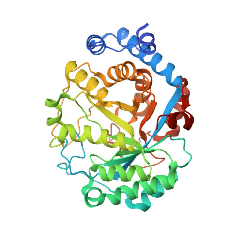
Crystal structure of methylornithine synthase (PylB): insights into the pyrrolysine biosynthesis.

Quitterer, F.List, A.Eisenreich, W.Bacher, A.Groll, M.

(2012) Angew Chem Int Ed Engl 51: 1339-1342


Macromolecules
Find similar proteins by:  (by identity cutoff)  |  3D Structure
Entity ID: 1
MoleculeChains Sequence LengthOrganismDetailsImage
methylornithine synthase PylB350Methanosarcina barkeri str. FusaroMutation(s): 0 
Gene Names: Mbar_A0838
EC: 2.8.1.6
UniProt
Find proteins for Q46E78 (Methanosarcina barkeri (strain Fusaro / DSM 804))
Explore Q46E78 
Go to UniProtKB:  Q46E78
Entity Groups  
Sequence Clusters30% Identity50% Identity70% Identity90% Identity95% Identity100% Identity
UniProt GroupQ46E78
Sequence Annotations
Expand
  • Reference Sequence
Experimental Data & Validation

Experimental Data

  • Method: X-RAY DIFFRACTION
  • Resolution: 1.50 Å
  • R-Value Free: 0.165 
  • R-Value Work: 0.129 
  • R-Value Observed: 0.130 
  • Space Group: P 32 2 1
Unit Cell:
Length ( Å )Angle ( ˚ )
a = 77.02α = 90
b = 77.02β = 90
c = 104.95γ = 120
Software Package:
Software NamePurpose
XDSdata scaling
REFMACrefinement
XDSdata reduction
XSCALEdata scaling
REFMACphasing

Structure Validation

View Full Validation Report


Ligand Structure Quality Assessment 


Entry History 

Deposition Data

Revision History  (Full details and data files)

  • Version 1.0: 2011-11-30
    Type: Initial release
  • Version 1.1: 2012-04-18
    Changes: Database references
  • Version 1.2: 2024-02-28
    Changes: Data collection, Database references, Derived calculations