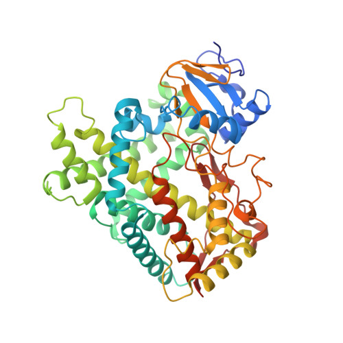
3T3S

Human Cytochrome P450 2A13 in complex with Pilocarpine


Experimental Data Snapshot

  • Method: X-RAY DIFFRACTION
  • Resolution: 3.00 Å
  • R-Value Free: 0.303 
  • R-Value Work: 0.213 
  • R-Value Observed: 0.218 

wwPDB Validation   3D Report Full Report


Ligand Structure Quality Assessment 


This is version 1.3 of the entry. See complete history


Literature

Structural comparison of cytochromes P450 2A6, 2A13, and 2E1 with pilocarpine.

DeVore, N.M.Meneely, K.M.Bart, A.G.Stephens, E.S.Battaile, K.P.Scott, E.E.

(2012) FEBS J 279: 1621-1631

  • DOI: https://doi.org/10.1111/j.1742-4658.2011.08412.x
  • Primary Citation of Related Structures:  
    3T3Q, 3T3R, 3T3S

  • PubMed Abstract: 

    Human xenobiotic-metabolizing cytochrome P450 (CYP) enzymes can each bind and monooxygenate a diverse set of substrates, including drugs, often producing a variety of metabolites. Additionally, a single ligand can interact with multiple CYP enzymes, but often the protein structural similarities and differences that mediate such overlapping selectivity are not well understood. Even though the CYP superfamily has a highly canonical global protein fold, there are large variations in the active site size, topology, and conformational flexibility. We have determined how a related set of three human CYP enzymes bind and interact with a common inhibitor, the muscarinic receptor agonist drug pilocarpine. Pilocarpine binds and inhibits the hepatic CYP2A6 and respiratory CYP2A13 enzymes much more efficiently than the hepatic CYP2E1 enzyme. To elucidate key residues involved in pilocarpine binding, crystal structures of CYP2A6 (2.4 Å), CYP2A13 (3.0 Å), CYP2E1 (2.35 Å), and the CYP2A6 mutant enzyme, CYP2A6 I208S/I300F/G301A/S369G (2.1 Å) have been determined with pilocarpine in the active site. In all four structures, pilocarpine coordinates to the heme iron, but comparisons reveal how individual residues lining the active sites of these three distinct human enzymes interact differently with the inhibitor pilocarpine.


  • Organizational Affiliation

    Department of Medicinal Chemistry, The University of Kansas, Lawrence, KS 66045, USA.


Macromolecules
Find similar proteins by:  (by identity cutoff)  |  3D Structure
Entity ID: 1
MoleculeChains Sequence LengthOrganismDetailsImage
Cytochrome P450 2A13
A, B, C, D, E
A, B, C, D, E, F, G, H
476Homo sapiensMutation(s): 0 
Gene Names: CYP2A13
EC: 1.14.14.1
UniProt & NIH Common Fund Data Resources
Find proteins for Q16696 (Homo sapiens)
Explore Q16696 
Go to UniProtKB:  Q16696
PHAROS:  Q16696
GTEx:  ENSG00000197838 
Entity Groups  
Sequence Clusters30% Identity50% Identity70% Identity90% Identity95% Identity100% Identity
UniProt GroupQ16696
Sequence Annotations
Expand
  • Reference Sequence
Small Molecules
Ligands 2 Unique
IDChains Name / Formula / InChI Key2D Diagram3D Interactions
HEM
Query on HEM

Download Ideal Coordinates CCD File 
I [auth A]
K [auth B]
M [auth C]
O [auth D]
Q [auth E]
I [auth A],
K [auth B],
M [auth C],
O [auth D],
Q [auth E],
S [auth F],
U [auth G],
V [auth H]
PROTOPORPHYRIN IX CONTAINING FE
C34 H32 Fe N4 O4
KABFMIBPWCXCRK-RGGAHWMASA-L
9PL
Query on 9PL

Download Ideal Coordinates CCD File 
J [auth A]
L [auth B]
N [auth C]
P [auth D]
R [auth E]
J [auth A],
L [auth B],
N [auth C],
P [auth D],
R [auth E],
T [auth F]
(3S,4R)-3-ethyl-4-[(1-methyl-1H-imidazol-5-yl)methyl]dihydrofuran-2(3H)-one
C11 H16 N2 O2
QCHFTSOMWOSFHM-WPRPVWTQSA-N
Binding Affinity Annotations 
IDSourceBinding Affinity
9PL BindingDB:  3T3S Ki: min: 1400, max: 3000 (nM) from 2 assay(s)
Kd: min: 3000, max: 3600 (nM) from 2 assay(s)
Experimental Data & Validation

Experimental Data

  • Method: X-RAY DIFFRACTION
  • Resolution: 3.00 Å
  • R-Value Free: 0.303 
  • R-Value Work: 0.213 
  • R-Value Observed: 0.218 
  • Space Group: P 1
Unit Cell:
Length ( Å )Angle ( ˚ )
a = 71.501α = 101.01
b = 119.862β = 101.72
c = 154.871γ = 93.59
Software Package:
Software NamePurpose
Blu-Icedata collection
PHASERphasing
REFMACrefinement
MOSFLMdata reduction
SCALAdata scaling

Structure Validation

View Full Validation Report


Ligand Structure Quality Assessment 


Entry History 

Deposition Data

Revision History  (Full details and data files)

  • Version 1.0: 2011-12-07
    Type: Initial release
  • Version 1.1: 2012-06-06
    Changes: Database references
  • Version 1.2: 2017-11-08
    Changes: Refinement description
  • Version 1.3: 2023-09-13
    Changes: Data collection, Database references, Derived calculations, Refinement description