Increased structural flexibility at the active site of a fluorophore-conjugated beta-lactamase distinctively impacts its binding toward diverse cephalosporin antibiotics
Wong, W.-T., Chan, K.-C., So, P.-K., Yap, H.-K., Chung, W.-H., Leung, Y.-C., Wong, K.-Y., Zhao, Y.-X.(2011) J Biol Chem 286: 31771-31780
- PubMed: 21705325
- DOI: https://doi.org/10.1074/jbc.M110.198895
- Primary Citation of Related Structures:
3SH7, 3SH8, 3SH9 - PubMed Abstract:
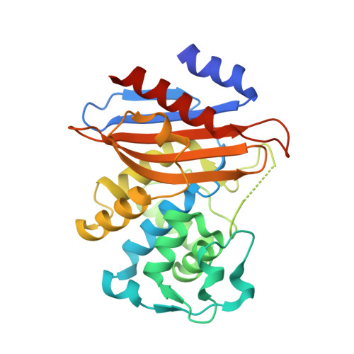
The Ω-loop at the active site of β-lactamases exerts significant impact on the kinetics and substrate profile of these enzymes by forming part of the substrate binding site and posing as steric hindrance toward bulky substrates. Mutating certain residues on the Ω-loop has been a general strategy for molecular evolution of β-lactamases to expand their hydrolytic activity toward extended-spectrum antibiotics through a mechanism believed to involve enhanced structural flexibility of the Ω-loop. Yet no structural information is available that demonstrates such flexibility or its relation to substrate profile and enzyme kinetics. Here we report an engineered β-lactamase that contains an environment-sensitive fluorophore conjugated near its active site to probe the structural dynamics of the Ω-loop and to detect the binding of diverse substrates. Our results show that this engineered β-lactamase has improved binding kinetics and positive fluorescence signal toward oxyimino-cephalosporins, but shows little such effect to non-oxyimino-cephalosporins. Structural studies reveal that the Ω-loop adopts a less stabilized structure, and readily undergoes conformational change to accommodate the binding of bulky oxyimino-cephalosporins while no such change is observed for non-oxyimino-cephalosporins. Mutational studies further confirm that this substrate-induced structural change is directly responsible for the positive fluorescence signal specific to oxyimino-cephalosporins. Our data provide mechanistic evidence to support the long-standing model that the evolutionary strategy of mutating the Ω-loop leads to increased structural flexibility of this region, which in turn facilitates the binding of extended spectrum β-lactam antibiotics. The oxyimino-cephalosporin-specific fluorescence profile of our engineered β-lactamase also demonstrates the possibility of designing substrate-selective biosensing systems.
Organizational Affiliation:
Department of Applied Biology and Chemical Technology, State Key Laboratory of Chirosciences, The Hong Kong Polytechnic University, Hung Hom, Kowloon, Hong Kong, China.