3S5U

Crystal structure of CRISPR associated protein


Experimental Data Snapshot

  • Method: X-RAY DIFFRACTION
  • Resolution: 2.70 Å
  • R-Value Free: 0.275 
  • R-Value Work: 0.219 
  • R-Value Observed: 0.219 

wwPDB Validation   3D Report Full Report


This is version 1.5 of the entry. See complete history


Literature

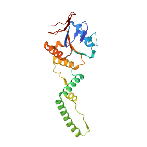
Crystal structure of clustered regularly interspaced short palindromic repeats (CRISPR)-associated Csn2 protein revealed Ca2+-dependent double-stranded DNA binding activity.

Nam, K.H.Kurinov, I.Ke, A.

(2011) J Biol Chem 286: 30759-30768

  • DOI: https://doi.org/10.1074/jbc.M111.256263
  • Primary Citation of Related Structures:  
    3S5U

  • PubMed Abstract: 

    Clustered regularly interspaced short palindromic repeats (CRISPR) and their associated protein genes (cas genes) are widespread in bacteria and archaea. They form a line of RNA-based immunity to eradicate invading bacteriophages and malicious plasmids. A key molecular event during this process is the acquisition of new spacers into the CRISPR loci to guide the selective degradation of the matching foreign genetic elements. Csn2 is a Nmeni subtype-specific cas gene required for new spacer acquisition. Here we characterize the Enterococcus faecalis Csn2 protein as a double-stranded (ds-) DNA-binding protein and report its 2.7 Å tetrameric ring structure. The inner circle of the Csn2 tetrameric ring is ∼26 Å wide and populated with conserved lysine residues poised for nonspecific interactions with ds-DNA. Each Csn2 protomer contains an α/β domain and an α-helical domain; significant hinge motion was observed between these two domains. Ca(2+) was located at strategic positions in the oligomerization interface. We further showed that removal of Ca(2+) ions altered the oligomerization state of Csn2, which in turn severely decreased its affinity for ds-DNA. In summary, our results provided the first insight into the function of the Csn2 protein in CRISPR adaptation by revealing that it is a ds-DNA-binding protein functioning at the quaternary structure level and regulated by Ca(2+) ions.


  • Organizational Affiliation

    Department of Molecular Biology and Genetics, Cornell University, Ithaca, New York 14850.


Macromolecules
Find similar proteins by:  (by identity cutoff)  |  3D Structure
Entity ID: 1
MoleculeChains Sequence LengthOrganismDetailsImage
Putative uncharacterized protein
A, B, C, D, E
A, B, C, D, E, F, G, H
220Enterococcus faecalis ATCC 4200Mutation(s): 0 
Gene Names: EFDG_01320
UniProt
Find proteins for C7UDU4 (Enterococcus faecalis)
Explore C7UDU4 
Go to UniProtKB:  C7UDU4
Entity Groups  
Sequence Clusters30% Identity50% Identity70% Identity90% Identity95% Identity100% Identity
UniProt GroupC7UDU4
Sequence Annotations
Expand
  • Reference Sequence
Small Molecules
Ligands 1 Unique
IDChains Name / Formula / InChI Key2D Diagram3D Interactions
CA
Query on CA

Download Ideal Coordinates CCD File 
I [auth A]
J [auth A]
K [auth B]
L [auth B]
M [auth C]
I [auth A],
J [auth A],
K [auth B],
L [auth B],
M [auth C],
N [auth C],
O [auth D],
P [auth D],
Q [auth E],
R [auth E],
S [auth F],
T [auth F],
U [auth G],
V [auth G],
W [auth H],
X [auth H]
CALCIUM ION
Ca
BHPQYMZQTOCNFJ-UHFFFAOYSA-N
Experimental Data & Validation

Experimental Data

  • Method: X-RAY DIFFRACTION
  • Resolution: 2.70 Å
  • R-Value Free: 0.275 
  • R-Value Work: 0.219 
  • R-Value Observed: 0.219 
  • Space Group: P 21 21 21
Unit Cell:
Length ( Å )Angle ( ˚ )
a = 104.112α = 90
b = 140.091β = 90
c = 148.434γ = 90
Software Package:
Software NamePurpose
PHENIXrefinement
PDB_EXTRACTdata extraction
HKL-2000data collection
HKL-2000data reduction
HKL-2000data scaling
SHELXSphasing
REFMACrefinement

Structure Validation

View Full Validation Report


Entry History 

Deposition Data

Revision History  (Full details and data files)

  • Version 1.0: 2011-06-22
    Type: Initial release
  • Version 1.1: 2011-07-13
    Changes: Version format compliance
  • Version 1.2: 2017-11-08
    Changes: Refinement description
  • Version 1.3: 2018-11-28
    Changes: Data collection, Database references
  • Version 1.4: 2019-07-17
    Changes: Data collection, Refinement description
  • Version 1.5: 2024-02-28
    Changes: Data collection, Database references, Derived calculations