3RQ4

Crystal structure of suppressor of variegation 4-20 homolog 2


Experimental Data Snapshot

  • Method: X-RAY DIFFRACTION
  • Resolution: 1.80 Å
  • R-Value Free: 0.221 
  • R-Value Work: 0.178 
  • R-Value Observed: 0.180 

wwPDB Validation   3D Report Full Report


Ligand Structure Quality Assessment 


This is version 1.4 of the entry. See complete history


Literature

Crystal structures of the human histone H4K20 methyltransferases SUV420H1 and SUV420H2.

Wu, H.Siarheyeva, A.Zeng, H.Lam, R.Dong, A.Wu, X.H.Li, Y.Schapira, M.Vedadi, M.Min, J.

(2013) FEBS Lett 587: 3859-3868

  • DOI: https://doi.org/10.1016/j.febslet.2013.10.020
  • Primary Citation of Related Structures:  
    3RQ4, 3S8P

  • PubMed Abstract: 

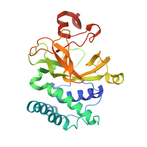
    SUV420H1 and SUV420H2 are two highly homologous enzymes that methylate lysine 20 of histone H4 (H4K20), a mark that has been implicated in transcriptional regulation. In this study, we present the high-resolution crystal structures of human SUV420H1 and SUV420H2 in complex with SAM, and report their substrate specificity. Both methyltransferases have a unique N-terminal domain and Zn-binding post-SET domain, and prefer the monomethylated histone H4K20 as a substrate in vitro. No histone H4K20 trimethylation activity was detected by our radioactivity-based assay for either enzyme, consistent with the presence of a conserved serine residue that forms a hydrogen bond with the target lysine side-chain and limits the methylation level.


Macromolecules
Find similar proteins by:  (by identity cutoff)  |  3D Structure
Entity ID: 1
MoleculeChains Sequence LengthOrganismDetailsImage
Histone-lysine N-methyltransferase SUV420H2247Homo sapiensMutation(s): 0 
Gene Names: SUV420H2KMT5CPP7130
EC: 2.1.1.43
UniProt & NIH Common Fund Data Resources
Find proteins for Q86Y97 (Homo sapiens)
Explore Q86Y97 
Go to UniProtKB:  Q86Y97
PHAROS:  Q86Y97
GTEx:  ENSG00000133247 
Entity Groups  
Sequence Clusters30% Identity50% Identity70% Identity90% Identity95% Identity100% Identity
UniProt GroupQ86Y97
Sequence Annotations
Expand
  • Reference Sequence
Small Molecules
Ligands 4 Unique
IDChains Name / Formula / InChI Key2D Diagram3D Interactions
SAM
Query on SAM

Download Ideal Coordinates CCD File 
C [auth A]S-ADENOSYLMETHIONINE
C15 H22 N6 O5 S
MEFKEPWMEQBLKI-FCKMPRQPSA-N
ZN
Query on ZN

Download Ideal Coordinates CCD File 
B [auth A]ZINC ION
Zn
PTFCDOFLOPIGGS-UHFFFAOYSA-N
EDO
Query on EDO

Download Ideal Coordinates CCD File 
D [auth A]1,2-ETHANEDIOL
C2 H6 O2
LYCAIKOWRPUZTN-UHFFFAOYSA-N
UNX
Query on UNX

Download Ideal Coordinates CCD File 
AA [auth A]
BA [auth A]
CA [auth A]
DA [auth A]
E [auth A]
AA [auth A],
BA [auth A],
CA [auth A],
DA [auth A],
E [auth A],
EA [auth A],
F [auth A],
FA [auth A],
G [auth A],
H [auth A],
I [auth A],
J [auth A],
K [auth A],
L [auth A],
M [auth A],
N [auth A],
O [auth A],
P [auth A],
Q [auth A],
R [auth A],
S [auth A],
T [auth A],
U [auth A],
V [auth A],
W [auth A],
X [auth A],
Y [auth A],
Z [auth A]
UNKNOWN ATOM OR ION
X
Experimental Data & Validation

Experimental Data

  • Method: X-RAY DIFFRACTION
  • Resolution: 1.80 Å
  • R-Value Free: 0.221 
  • R-Value Work: 0.178 
  • R-Value Observed: 0.180 
  • Space Group: P 21 21 21
Unit Cell:
Length ( Å )Angle ( ˚ )
a = 34.68α = 90
b = 60.151β = 90
c = 126.601γ = 90
Software Package:
Software NamePurpose
Blu-Icedata collection
MOLREPphasing
REFMACrefinement
Cootmodel building
HKL-2000data reduction
HKL-2000data scaling

Structure Validation

View Full Validation Report


Ligand Structure Quality Assessment 


Entry History 

Revision History  (Full details and data files)

  • Version 1.0: 2011-06-01
    Type: Initial release
  • Version 1.1: 2011-07-13
    Changes: Version format compliance
  • Version 1.2: 2014-01-15
    Changes: Database references
  • Version 1.3: 2024-02-28
    Changes: Data collection, Database references, Derived calculations
  • Version 1.4: 2024-04-03
    Changes: Refinement description