3RCC

Crystal Structure of the Streptococcus agalactiae Sortase A


Experimental Data Snapshot

  • Method: X-RAY DIFFRACTION
  • Resolution: 3.10 Å
  • R-Value Free: 0.299 
  • R-Value Work: 0.232 
  • R-Value Observed: 0.235 

wwPDB Validation   3D Report Full Report


This is version 1.2 of the entry. See complete history


Literature

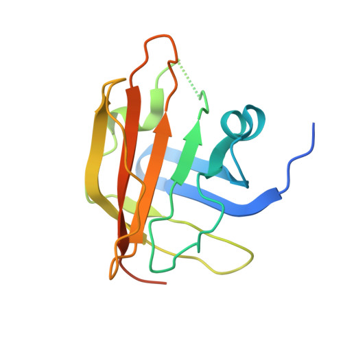
Structural differences between the Streptococcus agalactiae housekeeping and pilus-specific sortases: SrtA and SrtC1.

Khare, B.Krishnan, V.Rajashankar, K.R.I-Hsiu, H.Xin, M.Ton-That, H.Narayana, S.V.

(2011) PLoS One 6: e22995-e22995

  • DOI: https://doi.org/10.1371/journal.pone.0022995
  • Primary Citation of Related Structures:  
    3RBI, 3RBJ, 3RBK, 3RCC

  • PubMed Abstract: 

    The assembly of pili on the cell wall of Gram-positive bacteria requires transpeptidase enzymes called sortases. In Streptococcus agalactiae, the PI-1 pilus island of strain 2603V/R encodes two pilus-specific sortases (SrtC1 and SrtC2) and three pilins (GBS80, GBS52 and GBS104). Although either pilus-specific sortase is sufficient for the polymerization of the major pilin, GBS80, incorporation of the minor pilins GBS52 and GBS104 into the pilus structure requires SrtC1 and SrtC2, respectively. The S. agalactiae housekeeping sortase, SrtA, whose gene is present at a different location and does not catalyze pilus polymerization, was shown to be involved in cell wall anchoring of pilus polymers. To understand the structural basis of sortases involved in such diverse functions, we determined the crystal structures of S. agalactiae SrtC1 and SrtA. Both enzymes are made of an eight-stranded beta-barrel core with variations in their active site architecture. SrtA exhibits a catalytic triad arrangement similar to that in Streptococcus pyogenes SrtA but different from that in Staphylococcus aureus SrtA. In contrast, the SrtC1 enzyme contains an N-terminal helical domain and a 'lid' in its putative active site, which is similar to that seen in Streptococcus pneumoniae pilus-specific sortases, although with subtle differences in positioning and composition. To understand the effect of such differences on substrate recognition, we have also determined the crystal structure of a SrtC1 mutant, in which the conserved DP(W/F/Y) motif was replaced with the sorting signal motif of GBS80, IPNTG. By comparing the structures of WT wild type SrtA and SrtC1 and the 'lid' mutant of SrtC1, we propose that structural elements within the active site and the lid may be important for defining the role of specific sortase in pili biogenesis.


  • Organizational Affiliation

    Center for Biophysical Sciences and Engineering, University of Alabama at Birmingham, Birmingham, Alabama, United States of America.


Macromolecules
Find similar proteins by:  (by identity cutoff)  |  3D Structure
Entity ID: 1
MoleculeChains Sequence LengthOrganismDetailsImage
Sortase SrtA
A, B, C, D, E
A, B, C, D, E, F, G, H, I, J, K, L, M, N, O, P, Q, R
160Streptococcus agalactiae serogroup VMutation(s): 0 
Gene Names: SAG0961srtAsrtA (SAG0961)
UniProt
Find proteins for Q8DZY1 (Streptococcus agalactiae serotype V (strain ATCC BAA-611 / 2603 V/R))
Explore Q8DZY1 
Go to UniProtKB:  Q8DZY1
Entity Groups  
Sequence Clusters30% Identity50% Identity70% Identity90% Identity95% Identity100% Identity
UniProt GroupQ8DZY1
Sequence Annotations
Expand
  • Reference Sequence
Small Molecules
Ligands 1 Unique
IDChains Name / Formula / InChI Key2D Diagram3D Interactions
ZN
Query on ZN

Download Ideal Coordinates CCD File 
AA [auth B]
AB [auth H]
AC [auth O]
BA [auth B]
BB [auth H]
AA [auth B],
AB [auth H],
AC [auth O],
BA [auth B],
BB [auth H],
BC [auth O],
CA [auth B],
CB [auth H],
CC [auth O],
DA [auth C],
DB [auth I],
DC [auth P],
EA [auth C],
EB [auth I],
EC [auth P],
FA [auth C],
FB [auth I],
FC [auth P],
GA [auth C],
GB [auth I],
GC [auth P],
HA [auth C],
HB [auth J],
HC [auth Q],
IA [auth D],
IB [auth J],
IC [auth Q],
JA [auth D],
JB [auth J],
JC [auth Q],
KA [auth D],
KB [auth K],
KC [auth Q],
LA [auth D],
LB [auth K],
LC [auth R],
MA [auth E],
MB [auth K],
MC [auth R],
NA [auth E],
NB [auth L],
OA [auth E],
OB [auth L],
PA [auth E],
PB [auth M],
QA [auth F],
QB [auth M],
RA [auth F],
RB [auth M],
S [auth A],
SA [auth G],
SB [auth M],
T [auth A],
TA [auth G],
TB [auth M],
U [auth A],
UA [auth G],
UB [auth M],
V [auth A],
VA [auth G],
VB [auth N],
W [auth A],
WA [auth G],
WB [auth N],
X [auth B],
XA [auth G],
XB [auth N],
Y [auth B],
YA [auth G],
YB [auth N],
Z [auth B],
ZA [auth H],
ZB [auth O]
ZINC ION
Zn
PTFCDOFLOPIGGS-UHFFFAOYSA-N
Experimental Data & Validation

Experimental Data

  • Method: X-RAY DIFFRACTION
  • Resolution: 3.10 Å
  • R-Value Free: 0.299 
  • R-Value Work: 0.232 
  • R-Value Observed: 0.235 
  • Space Group: C 1 2 1
Unit Cell:
Length ( Å )Angle ( ˚ )
a = 237.674α = 90
b = 167.759β = 93.55
c = 97.536γ = 90
Software Package:
Software NamePurpose
HKL-2000data collection
PHASERphasing
REFMACrefinement
HKL-2000data reduction
HKL-2000data scaling

Structure Validation

View Full Validation Report


Entry History 

Deposition Data

Revision History  (Full details and data files)

  • Version 1.0: 2011-09-07
    Type: Initial release
  • Version 1.1: 2012-03-28
    Changes: Database references
  • Version 1.2: 2024-02-21
    Changes: Data collection, Database references, Derived calculations