3R7O

Structure of dually phosphorylated c-MET receptor kinase in complex with an MK-2461 analog


Experimental Data Snapshot

  • Method: X-RAY DIFFRACTION
  • Resolution: 2.30 Å
  • R-Value Free: 0.240 
  • R-Value Work: 0.211 
  • R-Value Observed: 0.213 

wwPDB Validation   3D Report Full Report


Ligand Structure Quality Assessment 


This is version 1.2 of the entry. See complete history


Literature

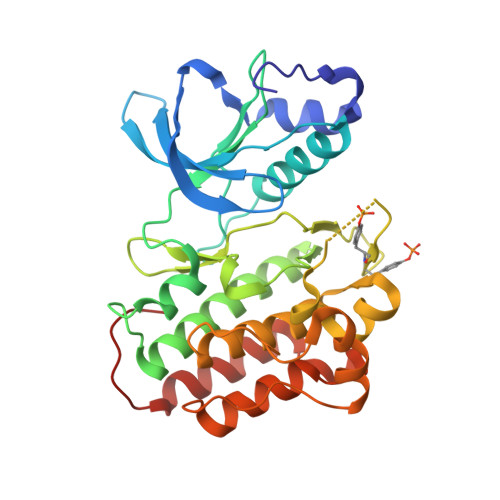
Structural basis for selective small molecule kinase inhibition of activated c-Met.

Rickert, K.W.Patel, S.B.Allison, T.J.Byrne, N.J.Darke, P.L.Ford, R.E.Guerin, D.J.Hall, D.L.Kornienko, M.Lu, J.Munshi, S.K.Reid, J.C.Shipman, J.M.Stanton, E.F.Wilson, K.J.Young, J.R.Soisson, S.M.Lumb, K.J.

(2011) J Biol Chem 286: 11218-11225

  • DOI: https://doi.org/10.1074/jbc.M110.204404
  • Primary Citation of Related Structures:  
    3Q6U, 3Q6W, 3R7O

  • PubMed Abstract: 

    The receptor tyrosine kinase c-Met is implicated in oncogenesis and is the target for several small molecule and biologic agents in clinical trials for the treatment of cancer. Binding of the hepatocyte growth factor to the cell surface receptor of c-Met induces activation via autophosphorylation of the kinase domain. Here we describe the structural basis of c-Met activation upon autophosphorylation and the selective small molecule inhibiton of autophosphorylated c-Met. MK-2461 is a potent c-Met inhibitor that is selective for the phosphorylated state of the enzyme. Compound 1 is an MK-2461 analog with a 20-fold enthalpy-driven preference for the autophosphorylated over unphosphorylated c-Met kinase domain. The crystal structure of the unbound kinase domain phosphorylated at Tyr-1234 and Tyr-1235 shows that activation loop phosphorylation leads to the ejection and disorder of the activation loop and rearrangement of helix αC and the G loop to generate a viable active site. Helix αC adopts a orientation different from that seen in activation loop mutants. The crystal structure of the complex formed by the autophosphorylated c-Met kinase domain and compound 1 reveals a significant induced fit conformational change of the G loop and ordering of the activation loop, explaining the selectivity of compound 1 for the autophosphorylated state. The results highlight the role of structural plasticity within the kinase domain in imparting the specificity of ligand binding and provide the framework for structure-guided design of activated c-Met inhibitors.


  • Organizational Affiliation

    Global Structural Biology, Merck Research Laboratories, West Point, Pennsylvania 19486, USA.


Macromolecules
Find similar proteins by:  (by identity cutoff)  |  3D Structure
Entity ID: 1
MoleculeChains Sequence LengthOrganismDetailsImage
Hepatocyte growth factor receptor307Homo sapiensMutation(s): 0 
Gene Names: MET
EC: 2.7.10.1
UniProt & NIH Common Fund Data Resources
Find proteins for P08581 (Homo sapiens)
Explore P08581 
Go to UniProtKB:  P08581
PHAROS:  P08581
GTEx:  ENSG00000105976 
Entity Groups  
Sequence Clusters30% Identity50% Identity70% Identity90% Identity95% Identity100% Identity
UniProt GroupP08581
Sequence Annotations
Expand
  • Reference Sequence
Small Molecules
Ligands 1 Unique
IDChains Name / Formula / InChI Key2D Diagram3D Interactions
M61
Query on M61

Download Ideal Coordinates CCD File 
B [auth A]N-[(2R)-1,4-dioxan-2-ylmethyl]-N-methyl-N'-{5-oxo-3-[1-(piperidin-4-yl)-1H-pyrazol-4-yl]-5H-benzo[4,5]cyclohepta[1,2-b]pyridin-7-yl}sulfuric diamide
C28 H32 N6 O5 S
IVWIPVHBADPMSP-XMMPIXPASA-N
Modified Residues  1 Unique
IDChains TypeFormula2D DiagramParent
PTR
Query on PTR
A
L-PEPTIDE LINKINGC9 H12 N O6 PTYR
Binding Affinity Annotations 
IDSourceBinding Affinity
M61 PDBBind:  3R7O Kd: 4.4 (nM) from 1 assay(s)
BindingDB:  3R7O IC50: min: 2.6, max: 3 (nM) from 2 assay(s)
Experimental Data & Validation

Experimental Data

  • Method: X-RAY DIFFRACTION
  • Resolution: 2.30 Å
  • R-Value Free: 0.240 
  • R-Value Work: 0.211 
  • R-Value Observed: 0.213 
  • Space Group: P 21 21 21
Unit Cell:
Length ( Å )Angle ( ˚ )
a = 42.671α = 90
b = 64.875β = 90
c = 110.99γ = 90
Software Package:
Software NamePurpose
DENZOdata reduction
SCALEPACKdata scaling
MOLREPphasing
BUSTER-TNTrefinement
PDB_EXTRACTdata extraction
ADSCdata collection
HKL-2000data reduction
BUSTERrefinement

Structure Validation

View Full Validation Report


Ligand Structure Quality Assessment 


Entry History 

Deposition Data

Revision History  (Full details and data files)

  • Version 1.0: 2012-02-01
    Type: Initial release
  • Version 1.1: 2013-01-09
    Changes: Non-polymer description, Structure summary
  • Version 1.2: 2017-11-08
    Changes: Refinement description