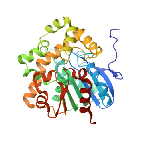
Mapping the reaction coordinates of enzymatic defluorination.
Chan, P.W., Yakunin, A.F., Edwards, E.A., Pai, E.F.(2011) J Am Chem Soc 133: 7461-7468
- PubMed: 21510690
- DOI: https://doi.org/10.1021/ja200277d
- Primary Citation of Related Structures:
3R3U, 3R3V, 3R3W, 3R3X, 3R3Y, 3R3Z, 3R40, 3R41 - PubMed Abstract:
The carbon-fluorine bond is the strongest covalent bond in organic chemistry, yet fluoroacetate dehalogenases can readily hydrolyze this bond under mild physiological conditions. Elucidating the molecular basis of this rare biocatalytic activity will provide the fundamental chemical insights into how this formidable feat is achieved. Here, we present a series of high-resolution (1.15-1.80 Å) crystal structures of a fluoroacetate dehalogenase, capturing snapshots along the defluorination reaction: the free enzyme, enzyme-fluoroacetate Michaelis complex, glycolyl-enzyme covalent intermediate, and enzyme-product complex. We demonstrate that enzymatic defluorination requires a halide pocket that not only supplies three hydrogen bonds to stabilize the fluoride ion but also is finely tailored for the smaller fluorine halogen atom to establish selectivity toward fluorinated substrates. We have further uncovered dynamics near the active site which may play pivotal roles in enzymatic defluorination. These findings may ultimately lead to the development of novel defluorinases that will enable the biotransformation of more complex fluorinated organic compounds, which in turn will assist the synthesis, detoxification, biodegradation, disposal, recycling, and regulatory strategies for the growing markets of organofluorines across major industrial sectors.
Organizational Affiliation:
Department of Biochemistry, University of Toronto, Toronto, Ontario M5S 1A8, Canada.